CORRECT DIAGNOSIS:
Sarcoidosis
DISCUSSION:
Sarcoidosis is a chronic, multisystem disorder of unknown cause characterized in affected organs by an accumulation of T lymphocytes and mononuclear phagocytes, noncaseating epitheliod granulomas, and derangements of the normal tissue architecture. Although there is usually skin anergy and depressed cellular immune process in the blood, Sarcoidosis is characterized at the sites of disease by exaggerated T helper lymphocyte immune processes. All parts of the body can be affected, but the organ most frequently affected in the lung. The involvement of the skin, eye, and lymph nodes is also common. The disease is often acute or subacute and self-limiting, but in many individuals it is chronic, waxing, and waning over several years.
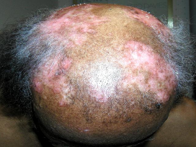
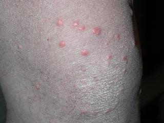
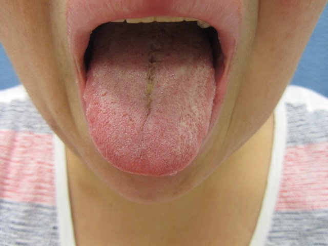
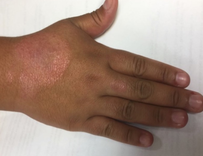
Cutaneous involvement in sarcoidosis may be classified as specific, which reveals granulomas on biopsy, or nonspecific, which is mainly reactive, such as erythema nodosum. The morphology of the lesions in sarcoidosis might include papules, nodules, plaques, subcutaneous nodule, scar sarcoidosis, erythroderma, ulceration, and verrucose, ichthyosiform, hypomelanotic, psoriasiform, and alopecia. Sarcoid, like syphilis, is a great mimic of other skin diseases and should be considered in the differential diagnosis of many different skin disorders. Sarcoidosis is a relatively common disease affecting individuals of both sexes and almost all ages, races, and geographic locations. Females appear slightly more susceptible than males. In the United States, the majority of the patients are African American, whereas in Europe he disease affects mostly Caucasians.
In most cases of sarcoidosis, the epidermis is normal or slightly atrophic. Exceptions occur in variants of verrucous sarcoid where there are acanthosis and hyperkeratoses; the latter is often seen in the ichthyosiform variant. Granulomas, the hallmark of sarcoidosis, is seen in many forms throughout the dermis, with the location depending on the type of cutaneous lesion. Granulomas variably contain necrosis, although not often, or fibrosis, or granular material. Granulomas classically are surrounded by modest lymphocytic infiltrate at the periphery making the “naked granuloma”. Large and irregular shaped giant cells may be seen in the lesions of sarcoidosis, and variably contain asteroid and Schaumann bodies. These bodies are also seen in other granulomatous disease processes such as tuberculosis. The asteroid bodies, more common that the Schaumann bodies in sarcoidosis, may be formed by trapped collagen bundles and when stained show a star-shaped eosinophilic structure. Schaumann bodies contain calcium crystals, are round and oval in shape, and are found more often in sarcoidosis that in tuberculosis.
For a definitive diagnosis of sarcoidosis, a biopsy is required, most commonly from the lung where the granulomatous process is most commonly found. In most circumstances, the diagnosis of sarcoidosis is a combination of physical, histological, and radiographic findings, although without consistent blood findings, diagnostic chest x-rays or scans(gallium 67), sarcoidosis is often confused with many other granulomatous disorders. Although not widely available, a helpful skin anergy test used is the Kviem-Siltzbach test. The test consists of an intradermal injection of heat stabilized antigen of sarcoidosis which is biopsied 4-6 weeks later for evaluation of the development of a granuloma. The test yields sarcoid-like granulomas in 70 to 80% of individuals with sarcoidosis and have less than a 5% false-positive reaction. Angiotensin-converting enzyme (ACE) levels may be elevated in all granulomatous diseases, including sarcoidosis. An elevated ACE level is suggestive but not diagnostic for granulomatous inflammation. The abnormal ACE level does not rule out sarcoidosis. If elevated, ACE levels may be used to monitor the activity of the disease.
TREATMENT:
Since most skin lesions of sarcoidosis are asymptomatic the major indication for treatment is cosmetic, ( except for disfiguring facial lesions). Systemic corticosteroids are virtually always beneficial cutaneous sarcoidosis. Unfortunately, the dose required to control the cutaneous disease may be too high to be ideal for long term use. Intralesional triamcinolone acetonide suspension 2.5mg/ml to 5.0mg/ml is very effective. For thinner lesions, super-potent topical steroids may be effective. Antimalarials chloroquine and hydroxychloroquine, and methotrexate have also shown partial or complete response.
This patient was treated with topical 0.1% triamcinolone cream BID for 3 weeks and had a resolution of erythema and a marked decrease in the number of lesions.
REFERENCES:
Odom, R. B. (2000). Andrews’ diseases of the skin (9th ed.). W.B. Saunders Company.
Elder, D. (1999). Synopsis and atlas of Lever’s histopathology of the skin. Lippincott Williams & Wilkins.
Fauci, A. S. (1998). Harrison’s principles of internal medicine (14th ed.). McGraw-Hill.


