CORRECT DIAGNOSIS:
Lichen myxedematosus
DISCUSSION:
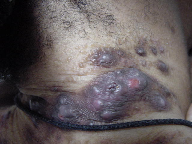
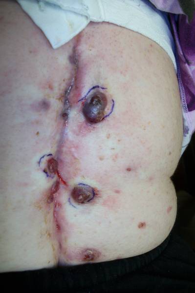
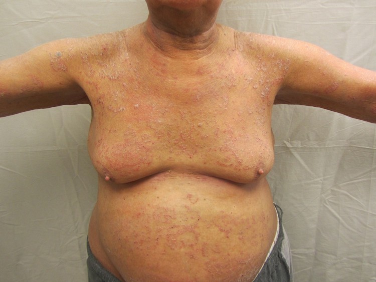
Lichen myxedematosus, localized form, affects adults of both sexes and appears from ages 30 – 80. The primary lesions are multiple, waxy, 2-mm to 4-mm dome-shaped or flat-topped papules that may coalesce into plaques or arrange in a linear arrangement. Other forms such as urticarial nodular annular lesions are not commonly seen. This commonly occurs on the dorsal hands, face, elbows, and extensor extremities. Pruritus may occur. Leonine facies may develop due to the coalescence of lesions on the forehead and glabella. The disease course is chronic and usually progressive. Treatment for the cutaneous form is ineffective.
The pathogenesis for this disease is unknown. It is associated with paraproteinemia that consists of myeloma-like homogenous serum globulinemia of IgG type with a predominately lambda light chain. The association with multiple myeloma is rare but it is suggested that papular mucinosis represents plasma cell dyscrasia. Scleromyxedema, a variant, the skin shows erythematous scleroderma-like induration accompanied by the rigidity of the lips, hands, arms, and legs. It is also associated with systemic manifestations such as severe proximal myopathy, inflammatory polyarthritis, central nervous system symptoms, acute organic brain syndrome, esophageal parasols, and hoarseness.
Pathology shows striking changes in the epidermis which show a horizontal band of mucinous material between collagen bundles. This material is glycosaminoglycan that stains with LCM blue at pH 2.5 and is susceptible to hyaluronidase. There is an increased number of fibroblasts, which appear plump and stellate, and dermatofibrosis. Clinically, the differential diagnosis should include scleredema, scleroderma, amyloidosus, disseminated granuloma annulare, malignant lymphoma, and dermatomyositis. The more localized form should be differentiated from colloid degeneration, lichen planus, morbus moniliformis and epithelioma adenoides cysticum.
TREATMENT:
The actual treatment for this patient: Intralesional Kenalog 5-mg per cc at monthly intervals with mild improvement. Other treatment options for cutaneous involvement include isotretinoin and etretinate, which have been associated with improvement. Alpha interferon, cyclosporin, PUVA, electron beam treatment, and dermabrasion are additional treatment options. Most of these treatments remain unsatisfactory.
REFERENCES:
Odom, R. B. (2000). Andrews’ diseases of the skin (9th ed.). W.B. Saunders Company.
Elder, D. (1997). Lever’s histopathology of the skin (8th ed.). Lippincott.
Fleischmajer, R. (Eds.). (1999). Dermatology and general medicine (5th ed.). McGraw-Hill Companies.


