Presenter: Laurie Schaeffer, D.O., Michael Eyre, D.O., Wendy McFalda, D.O., Cindy Lavery, D.O.
Dermatology Program: Pontiac Osteopathic Hospital
Program Director: Sandy Goldman, D.O.
Submitted on: Sep 29, 2002
CHIEF COMPLAINT: Pruritic, painful groin rash for approximately six weeks
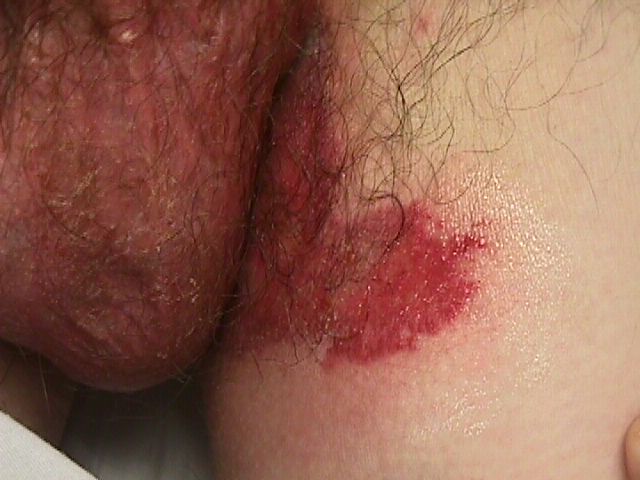
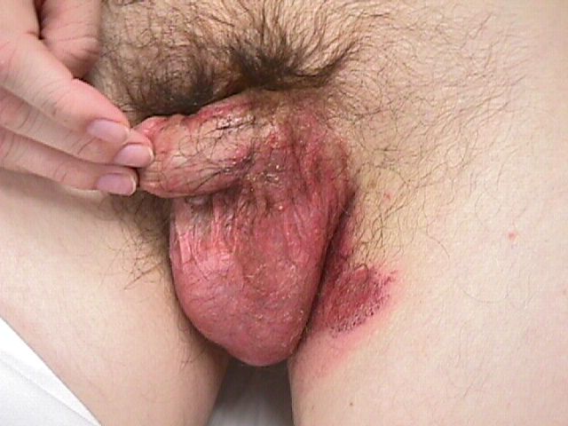
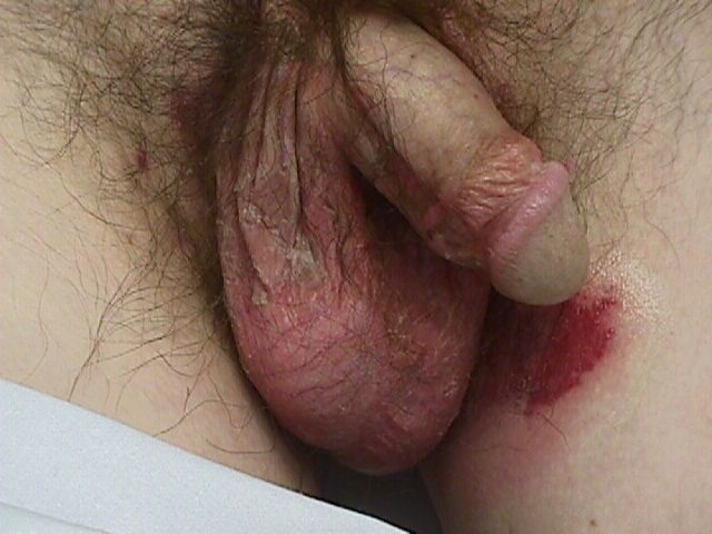
CLINICAL HISTORY: The patient presented with a painful, pruritic, and progressively worsening rash in the groin area. Initially starting on the sides of his scrotum, the rash had expanded to involve the entire scrotum, the sides of the penis, and the upper thighs. This condition severely impacted his ability to ambulate and maintain hygiene due to intense pain. He denied experiencing dysuria, hematuria, or any discharge, and could not recall having similar outbreaks in the past. Additionally, he reported no constitutional symptoms but mentioned occasional diarrhea.
In terms of previous treatment, the patient had applied Lotrisone cream, Mentax cream, and over-the-counter hydrocortisone creams with little relief.
The patient had been residing in a psychiatric group home for the past three months, during which he was prescribed Remeron, Wellbutrin, and Zyprexa, all initiated around the same time as his symptoms began. Recently, Trazodone was added to his medication regimen within the last week. Notably, the patient shared that he had undergone chemotherapy for a liver tumor, which is currently in remission.
PHYSICAL EXAM:
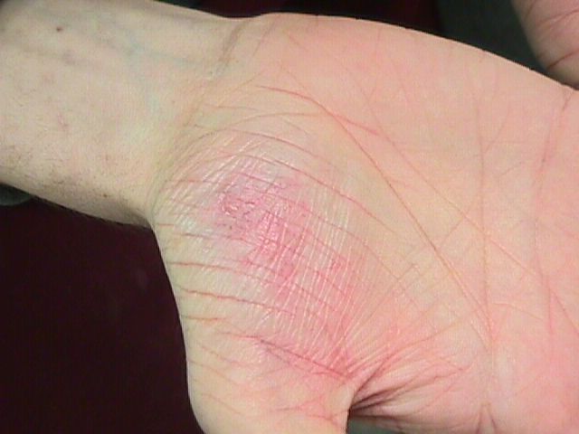
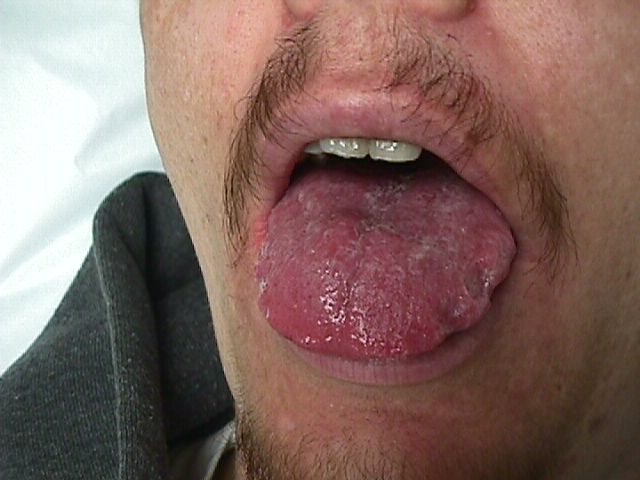
Physical exam revealed a 34-year-old cachectic white male with a flat affect. The patient had diffuse erythema with scale on the entire scalp. The scalp hair appeared thinned and brittle. The eyebrows, nasolabial folds, and perioral region displayed faint erythematous patches with light scale. The tongue was erythematous and enlarged with lateral imprints of dentition. Bilateral oral commissures displayed erythema and fissuring. No nail changes were noted. Beefy red, slightly erosive patches with surrounding erythema and scale were noted on the scrotum, base of the penis, and bilateral groin. The palms were noted to have erythema and scale with increased skin markings. Vesicles or bullae were absent.


LABORATORY TESTS:
Complete blood count revealed a normochromic/normocytic anemia. ALT and alkaline phosphatase were elevated at 74 (nl 7-40 IU/L) and 195 (nl 37-107 U/L), respectively. Intact PTH and serum calcium were normal. Serum zinc was normal at 1014 (nl 600-1200 UG/L). Insulin was elevated at 27.5 (nl 1-19 UU/ML). Gastrin was elevated at 112 (nl 0-90 PG/ML). Glucagon was elevated at 1650 (nl 40-130 NG/L).
CT of the Abdomen revealed calcification of the tail of the pancreas with multiple areas of metastatic lesions in the liver.
DERMATOHISTOPATHOLOGY:
Microscopic description: Histological specimens revealed psoriasiform hyperplasia with a moderate neutrophilic infiltrate. Dyskeratotic cells with cytoplasmic eosinophilic homogenization were noted in the upper epidermis along with marked hydropic swelling. Candida was also present.
DIFFERENTIAL DIAGNOSIS:
1. Acrodermatitis Enteropathica
2. Seborrheic Dermatitis
3. Necrolytic Migratory Erythema
4. Subcorneal Pustular Dermatosis

