CORRECT DIAGNOSIS:
Atypical fibroxanthoma
DISCUSSION:
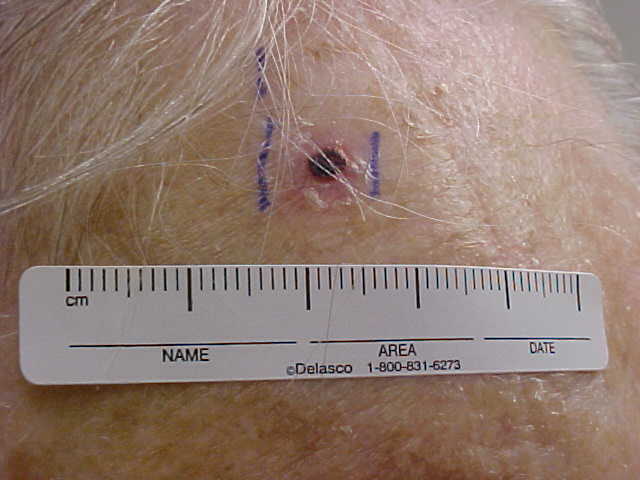
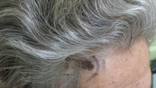
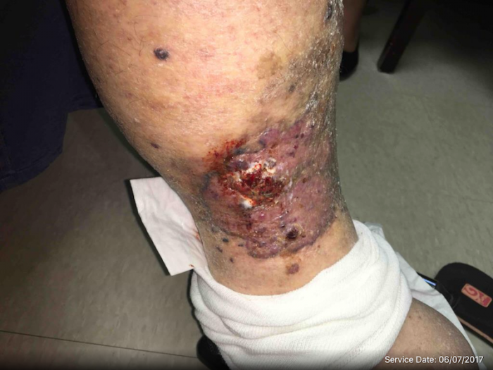
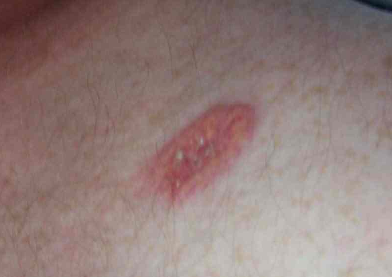
Atypical fibroxanthoma is a tumor that occurs in older patients in the areas of sun exposure and/or therapeutic radiation. The lesions clinically are suggestive of malignancy because they are fast-growing. This disease entity often leads to misdiagnosis and results in unnecessary and extensive surgery. It is a low-grade malignancy related to malignant fibrous histiocytoma. Because atypical fibroxanthoma is small in size and more superficially located, it has a much better prognosis than malignant fibrous histiocytoma. Some cases may represent primary squamous cell carcinoma (SCC) that fails to express keratin. The tumor is often presenting as a small, firm nodule with an eroded crusted surface. Histologically, lesions show a highly atypical and pleomorphic cellular appearance. The tumor consists of spindle cells mingled with atypical histiocytes. Vesicular nuclei are located in some spindle cells. The cytoplasm may be vacuolated and resemble the foamy cells of xanthomas.
They typically respond to simple excision but have a high rate of local recurrence. For this reason, Mohs surgery is the treatment of choice. Factors important to consider are lesion location, patient age, histopathologic appearance, and the observation that the tumor arises from the dermis, not the fat. Metastasis is rare.
TREATMENT:
For our patient, the lesion was excised with a specimen sent to pathology for confirmation. The margins were cleared and the patient was instructed to follow up for recheck in 3 months.
Evidence is accumulating that demonstrates that Mohs micrographic surgery, with its high reliability of complete tumor removal and tissue-conserving property, may be the treatment of choice for atypical fibroxanthoma on certain areas of the head and neck.
REFERENCES:
Lee, C. S., & Chou, S. T. (1998). p53 protein immunoreactivity in fibrohistiocytic tumors of the skin. Pathology, 30(3), 272–275. https://doi.org/10.1080/003130298100687 [PMID: 9739115]
Leong, A. S., & Milios, J. (1987). Atypical fibroxanthoma of the skin: A clinicopathological and immunohistochemical study and a discussion of its histogenesis. Histopathology, 11(5), 463–475. https://doi.org/10.1111/j.1365-2559.1987.tb02062.x [PMID: 3651780]
Ma, C. K., Zarbo, R. J., & Gown, A. M. (1992). Immunohistochemical characterization of atypical fibroxanthoma and dermatofibrosarcoma protuberans. American Journal of Clinical Pathology, 97(4), 478–483. https://doi.org/10.1093/ajcp/97.4.478 [PMID: 1555015]
Requena, L., Sangueza, O. P., Sanchez Yus, E., & Furio, V. (1997). Clear-cell atypical fibroxanthoma: An uncommon histopathologic variant of atypical fibroxanthoma. Journal of Cutaneous Pathology, 24(3), 176–182. https://doi.org/10.1111/j.1600-0560.1997.tb00573.x [PMID: 9141504]
Silvis, N. G., Swanson, P. E., Manivel, J. C., et al. (1988). Spindle-cell and pleomorphic neoplasms of the skin: A clinicopathologic and immunohistochemical study of 30 cases, with emphasis on “atypical fibroxanthomas.” American Journal of Dermatopathology, 10(1), 9–19. https://doi.org/10.1097/00000372-198802000-00002 [PMID: 3341718]
Starink, T. H., Hausman, R., Van Delden, L., & Neering, H. (1977). Atypical fibroxanthoma of the skin: Presentation of 5 cases and a review of the literature. British Journal of Dermatology, 97(2), 167–177. https://doi.org/10.1111/j.1365-2133.1977.tb13961.x [PMID: 329165]


