Presenter: Suleman Bangash, DO, Chief Resident and Carissa Summa, DO, Second Year Resident
Dermatology Program: New York United Medical Center, New York
Program Director: Cindy Hoffman, DO, FAOCD
Submitted on: January 1, 2005
CHIEF COMPLAINT: Growth on the Left Foot
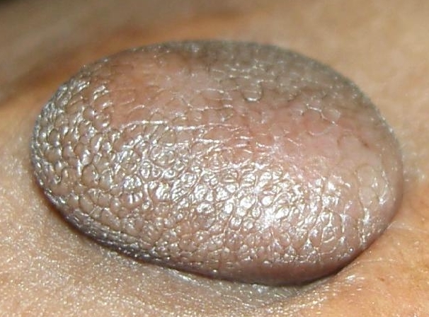
CLINICAL HISTORY: Patient with Milroy’s disease (congenital lymphedema) presented with a new growth on left foot that was slightly tender to palpation. She reported that the lesion began as a brown patch and slowly enlarged to a dome-shaped nodule over several years. She also reported multiple similar, but smaller lesions on her upper and lower extremities and trunk. The patient was only taking Hydrochlorothiazide for the lower extremity edema.
PHYSICAL EXAM:
A 3.0 X 3.2 cm, slightly hyperpigmented, soft, pedunculated nodule was present on the left dorsal foot. A total of nine hyperpigmented, firm 0.5 – 1.0 cm nodules were found on bilateral upper and lower extremities and trunk.
LABORATORY TESTS: N/A
DERMATOHISTOPATHOLOGY:
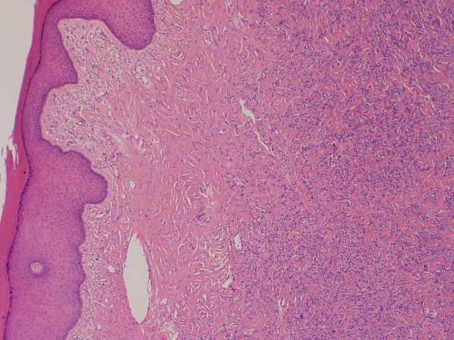
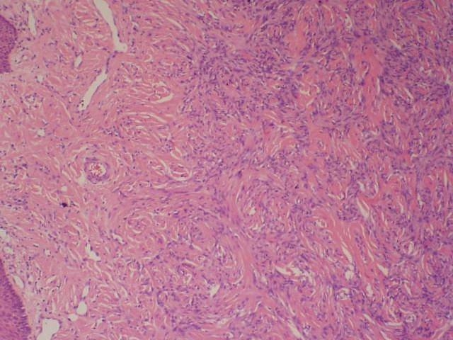
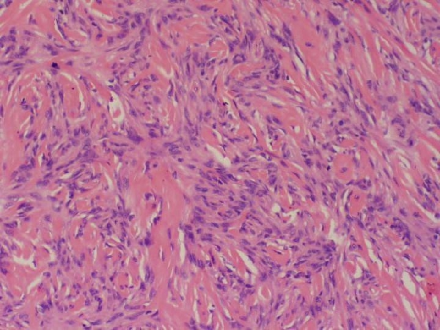
A shave biopsy of the left dorsal foot lesion was done.
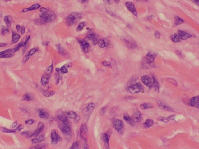
Spindle cell tumor composed of fibroblasts, histiocytes, and multinucleated giant cells in the dermis. The overlying epidermis is hyperplastic with elongation of rete ridges.
DIFFERENTIAL DIAGNOSIS:
1. Polypoid nodular dermatofibroma
2. Superficial leiomyoma
3. Keloid
4. Malignancy
5. Acquired Fibrokeratoma

