CORRECT DIAGNOSIS:
Systemic Lupus Erythematosus
DISCUSSION:
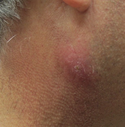
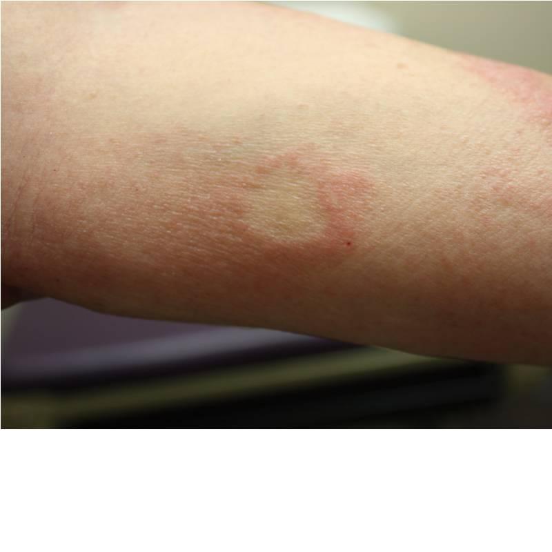
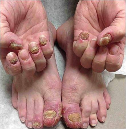
Our main differential was if this was a case of drug-induced lupus secondary to Lisinopril versus systemic lupus erythematosus. According to Litt, 9th edition, the rash is seen 1.5% of patients, and lupus erythematosus is listed as a reaction as well. However, the history of hypertension at the end of the pts third trimester of pregnancy without elevated liver functions, suggested that this disease process preceded the start of lisinopril. While the labs were pending we did hold the lisinopril and change to a B-blocker as an antihypertensive. In spite of this, at the next visit, two weeks later, she had more extensive skin involvement. The anti-histone antibodies were negative and this supported our suspicion that this was not drug-induced, but a true acute systemic lupus erythematosus.
Systemic lupus erythematosus affects one out of every 1,000 white persons and one out of every 250 black women from 18 to 65 years of age. The etiology still remains unclear however there is an abnormal production of antibodies by B cells. To make the diagnosis the patient must have four of eleven criteria developed by the American College of Rheumatology. Skin involvement occurs in 80% of cases. The earliest changes may be transitory or migratory arthralgias and fever, weight loss, pleuritis, adenopathy, or abdominal pain. Other abnormalities include thrombosis of various sized vessels, renal involvement (either nephritic or nephritic), myocarditis, pericarditis, Raynaud’s phenomenon, and CNS vasculitis, and ITP.
Our patient had 4 of the eleven criteria to make the diagnosis of SLE. She had the typical early arthralgias. She was also of the typical sex and age. She presented with preceding renal involvement with the history of hypertension in late pregnancy without the other typical “HELP?or preeclampsia findings. Her hypertension is likely secondary to this renal involvement as is her proteinuria. She was referred to a nephrologist and decided to follow up with her primary care physician. Unfortunately, she was lost to follow up. If she remained under our care we would have considered treating options with systemic corticosteroids, antimalarials, or other immunosuppressive agents. The literature supports using systemic steroids in cases with renal and central nervous system involvement, to effectively prolong survival.
TREATMENT:
Our patient was treated with Locoid cream BID to facial lesions, Cloderm cream BID to areas on arms and legs, and aggressive UVA/UVB sunblock. Sunblock, topical steroids, and antimalarials are important in the treatment of cutaneous lupus. Antimalarials used are hydroxychloroquine and quinacrine or chloroquine(has more of a risk of retinopathy). Oral steroids ( once the renal disease was better defined with at least a 24hr urine and possibly a kidney biopsy). Team approach- referral to a nephrologist and a rheumatologist or internist/ primary care physician. Oral NSAIDs are used for arthralgias; however, it is important to avoid in patients with nephritis and will increase the risk of gastric ulcers when used in combination with corticosteroids. Steroid sparing immunosuppressive medications like methotrexate or azathioprine can also be considered. Of note, thalidomide is one of the most effective drugs for the treatment of discoid lupus.
REFERENCES:
Litt, J. Z. (2003). Drug eruption reference manual (9th ed.). New York, NY: The Parthenon Publishing Group Inc.
Odom, R. B., James, W. D., & Berger, T. G. (2000). Andrews’ diseases of the skin. Philadelphia, PA: Saunders.
Petri, M. (1998). Treatment of systemic lupus erythematosus: An update. American Family Physician.


