CORRECT DIAGNOSIS:
Wegener’s Granulomatosis
DISCUSSION:
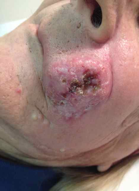
Wegener’s Granulomatosis, now known as Granulomatosis with Polyangiitis, is characterized by necrotizing granulomas in the upper and lower respiratory tracts, necrotizing angiitis of medium-sized blood vessels, and focal necrotizing glomerulitis. Common signs and symptoms include rhinorrhea, sinusitis, nasal and mucosal ulcerations, fever, weight loss, malaise, and “strawberry gums.” The condition typically presents in individuals aged 40 to 50, with a male-to-female ratio of 1.3:1. Nasal obstruction is frequent, and pulmonary nodules are observed in 40% to 70% of patients, which may ulcerate and bleed. Granulomas can also affect the ears and mouth, leading to tongue ulceration and perforating ulcers of the palate.
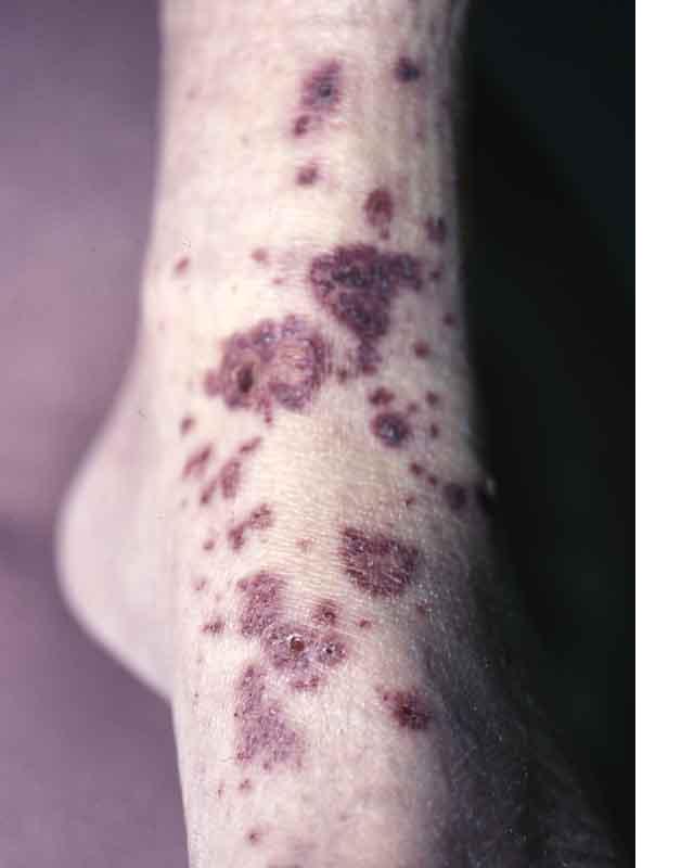
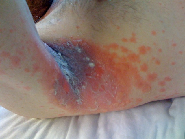
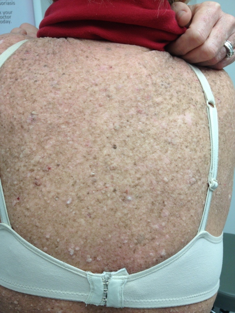
Additional findings may include tracheal stenosis and cutaneous manifestations in 45% of patients, with nodules that are firm, slightly tender, and flesh-colored before ulcerating. Necrotizing angiitis of the skin can present as petechiae, purpura, or pustular eruptions, while temporal arteritis-like symptoms and rare livedo reticularis may also occur. Focal necrotizing glomerulitis is found in 70% to 85% of cases, with renal failure being a common cause of death. Lung involvement is seen in 90% of patients, accompanied by arthralgia in 66%, eye involvement in 58%, CNS symptoms in 22%, and cardiac issues in 12%.
Radiographic findings can vary, with chest X-rays potentially being negative in up to 20% of cases, although they commonly show nodules, airspace opacities, atelectasis, reticular interstitial opacities, and adenopathy. CT scans typically reveal nodules and airspace consolidation, with sizes ranging from 5 mm to 10 cm. Airspace disease may be bilateral and diffuse due to pulmonary hemorrhage or localized with ill-defined margins and central cavitation. Interstitial abnormalities can include interlobular septal thickening, parenchymal bands, and bronchial wall thickening.
Histopathological examination typically shows leukocytoclastic vasculitis, sometimes with granulomatous inflammation. For diagnosis, C-ANCA is often positive but not specific to Wegener’s Granulomatosis. Lung or kidney biopsy, preferably lung biopsy, is crucial. Clinical criteria for diagnosis include nasal or oral inflammation, abnormal chest X-ray, urinary sediment abnormalities, and suggestive biopsy results, with a diagnosis confirmed if at least two out of four criteria are met, achieving 88% sensitivity and 92% specificity.
TREATMENT:
Our patient was initially started on intravenous Solu-Medrol and subsequently transitioned to cyclophosphamide. The treatment modalities included cyclophosphamide at a dosage of 2 mg/kg/day or a pulsed dose of 15 mg/kg every two weeks. Additional treatment options consisted of prednisone, azathioprine, cyclosporine, methotrexate (MTX), and Bactrim.
REFERENCES:
Bolognia, J. L., Jorizzo, J. L., Rapini, R. P., et al. (2003). Dermatology. Spain: Mosby.
Odom, R. B., & James, W. D. (2000). Andrews’ Diseases of the Skin. Philadelphia, PA: Elsevier.
Braunwald, E., Fauci, A. S., Kasper, D. L., et al. (2001). Harrison’s Principles of Internal Medicine. New York: McGraw-Hill.eMedicine.


