Presenter: Chris Weyer, DO; Bo Rivera, DO; Jonathan Cleaver, DO
Dermatology Program: Northeast Regional Medical Center – Kirksville
Program Director: Lloyd J. Cleaver, DO
Submitted on: March 14, 2010
CHIEF COMPLAINT: Oral and genital lesions
CLINICAL HISTORY: A 27-year-old female presents with a history of perioral lesions lasting at least one year and genital lesions persisting for over three years. She describes the genital lesions as itchy and burning, noting that they can bleed with excessive picking. Additionally, she reports experiencing burning sensations and increased frequency of urination. Despite recent normal pap smears, her symptoms have been concerning. Upon further questioning, the patient revealed a notable decrease in sweating. She has not received any prior treatments for her symptoms.
Her past medical history includes recurrent yeast infections following antibiotic use, and she is HIV negative. The patient has no significant surgical history. Socially, she smokes one pack of cigarettes per day and drinks alcohol rarely. Family history is unremarkable. Her current medications include ibuprofen as needed and a levonorgestrel intrauterine device (Mirena). She has no known allergies.
PHYSICAL EXAM:
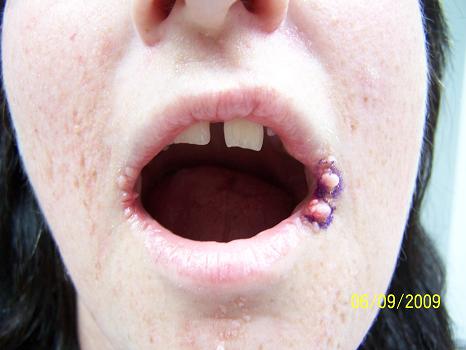
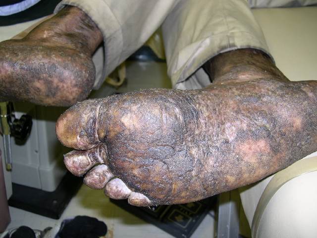
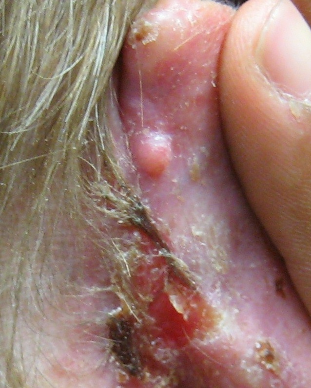
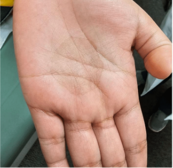
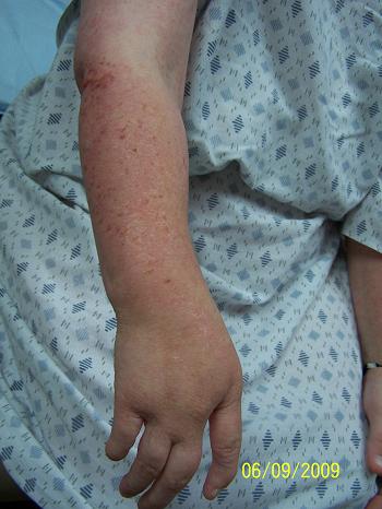
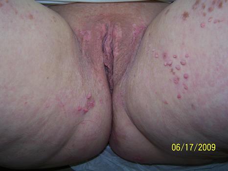
Examination revealed pink, verrucous papules, 2-4mm in size periorally, in the groin, on the upper medial thighs, and on the buttocks. Her bilateral arms and legs were significant for hypopigmented atrophic scars with telangiectasias and yellowish nodules in the linear array following Blaschko’s lines. Her right hand only had four digits and her right and left feet had three digits. Orally, poor dentition was noted.



LABORATORY TESTS:
X-ray examinations were reported as follows:
Bilateral femurs – There are longitudinal striations that involve the metaphysis with extensions into the epiphyses and diaphysis bilaterally. The findings are consistent with osteopathia striata.
Tibia/fibula – There are longitudinal striations that involve the metaphysis with extensions into the epiphyses and diaphysis bilaterally. The findings are consistent with osteopathia striata. There is an ill-defined radiolucent defect of the proximal right fibula, this may be consistent with an early chondroblastoma.
Right hand – The examination demonstrates a deformity of the right hand consistent with a cleft hand. There is a complete congenital absence of the fifth digit. Also noted are clinodactyly of the first digit, brachiadactyly of the fourth digit with shortening of the proximal phalanx and complete congenital absence of the middle phalanx. Additionally, the distal phalanx is rudimentary. There are congenital changes in the wrist as well. Marked enlargement of the capitate with congenital absence of the triquetrum and pisiform are seen. Findings suggesting acquired ulnar positive variance. Also, a 3.2 x 1.1 cm expansile radiolucent defect of the right second metacarpal is present. There are internal stippled calcifications with a surrounding sclerotic margin, possibly a solitary chondroblastoma.
Left hand – normal
DERMATOHISTOPATHOLOGY:
L lateral oral commissure: verrucous keratosis
L medial thigh: inflamed condyloma acuminatum
R superior lateral arm: dermal hypoplasia with normal elastin fibers demonstrated by elastin special stains
R labia majora – condyloma acuminatum
DIFFERENTIAL DIAGNOSIS:
1. Condyloma acuminatum
2. Nevus lipomatous superficialis
3. Goltz Syndrome
4. Incontinentia pigmentii
5. Rotmund-Thompson Syndrome

