CORRECT DIAGNOSIS:
Goltz Syndrome
DISCUSSION:
Goltz syndrome, or focal dermal hypoplasia, is a widespread dysplasia of mesodermal and ectodermal structures, with characteristic underdevelopment of the dermis10. Goltz syndrome was first described by Goltz in 1962. The main feature is profound dysplasia of connective tissue, especially the skin and bones4-7.
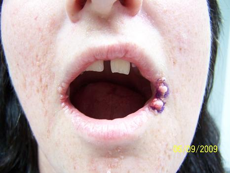
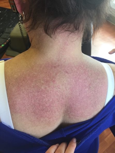
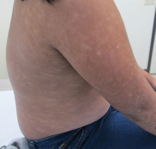
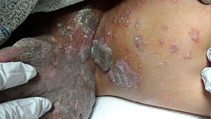
There are a wide variety of physical findings in Goltz syndrome. Numerous cutaneous manifestations of the disease exist and include linear, cribriform, or reticular areas of hypoplasia of the skin, striae distensae, verrucoid papillomas of the skin and mucous membranes, and soft yellowish nodules. The soft nodules represent the presence of fat in the dermis1. Atrophic macules appearing pink to red, telangiectasias, and raspberry-like papules are other cutaneous manifestations that may be present13. The raspberry-like papillomas can appear at any age. The most common locations are at the junction of mucosa and skin, lips, vulva, perianal, and periorbital areas. Alternate locations include the ears, fingers, toes, groin, umbilicus, oral, and esophagus3, 13. Many of these cutaneous lesions will follow the lines of Blaschko. The hair can have a sparse and brittle in appearance. When linear lesions become contiguous with the nails, nail dystrophy and anonychia may be present13. An early inflammatory vesicular eruption has been reported in some patients11.
Skeletal anomalies are also numerous, with the most common being syndactyly. The third and fourth digits on both the hands and the feet are often involved12. Polydactyly, adactyly, scoliosis, and hypoplasia of the distal extremities may also be found12. Approximately 80% of patients will have skeletal anomalies which may include dental defects such as dysplastic or absent teeth, enamel defects, and high arched or cleft palates14. Facial asymmetry, reduced bone density or osteoporosis, and fibrous dysplasia of the bone can be found2. Radiographic changes, osteopathia striata, may also be present and are characterized by asymptomatic, fine linear densities parallel to the axis of the long bones, but are not pathognomonic for Goltz syndrome6.
There are numerous other physical manifestations that are highly variable from patient to patient. Ocular involvement occurs 20% of the time and includes defects of the anterior chamber, coloboma of the iris, aniridia, choroidoretinal colobomata, microphthalmos, anophthalmos, asymmetry, and strabismus13. Renal involvement can present as horseshoe kidney or mild cystic dysplasia7. Multiple hernias have also been reported as complications including exomphalos, inguinal, and epigastric hernias. The intelligent quotient is usually normal in these patients; however, in severe cases, microcephaly and mental retardation can occur13.
Goltz syndrome has a variety of histopathologic findings. A normal epidermis is usually found overlying a hypoplastic, collagen scarce dermis14. The fatty herniations are consistent with the histologic findings of islands of adipose tissue found in the superficial dermis which impinges on the epidermis1. A narrow dermis is found along most of the dermal-epidermal junction and peri-appendageal areas, however, in areas with no dermis, ulceration presents and the epidermis is unable to survive14. Under electron microscopy, the dermis shows long filamentous strands that most likely represent immature collagen15. It is postulated that collagen synthesis is occurring at a normal rate, but is unable to form mature bundles13.
The genetic pattern for Goltz syndrome has been widely studied. The majority of cases have been in females, suggesting that there is an X-linked dominant inheritance pattern. This is consistent with male inheritors of the trait not being able to survive. Approximately 90 percent of cases have been female, with only approximately 30 cases being reported in males worldwide14. All of the male cases have been sporadic without any male-to-male transmission6. New research has found a link to the PORCN gene which is a regulator for Wnt signaling8,16.
Supportive therapy is the mainstay for treatment in Goltz syndrome. Reconstructive surgery is occasionally used. Cryotherapy has been studied for control of the papillomas associated with Goltz syndrome9. Most patients are able to live normal lives with normal lifespan.
TREATMENT:
In this patient, liquid nitrogen cryotherapy was used to manage the oral papillomas and liquid nitrogen cryotherapy along with podophyllin were utilized to manage the genital lesions.
REFERENCES:
Boente, M. del C., Asial, R. A., & Winik, B. C. (2007). Focal dermal hypoplasia: Ultrastructural abnormalities of the connective tissue. Journal of Cutaneous Pathology, 34(3), 181-187. https://doi.org/10.1111/j.1600-0560.2006.00534.x
Boothroyd, A. E., & Hall, C. M. (1988). The radiologic features of Goltz syndrome: Focal dermal hypoplasia: A report of two cases. Skeletal Radiology, 17(7), 505-508. https://doi.org/10.1007/BF00220563
Brinson, R. B., Schuman, B. M., & Mills, L. R. (1987). Multiple squamous papules of the oesophagus associated with Goltz syndrome. American Journal of Gastroenterology, 82(11), 1177-1179. https://doi.org/10.1111/j.1572-0241.1987.tb04596.x
Goltz, R. W. (1962). Focal dermal hypoplasia. Archives of Dermatology, 86(6), 708. https://doi.org/10.1001/archderm.1962.01580180036004
Goltz, R. W. (1990). Focal dermal hypoplasia. Pediatric Dermatology, 7(4), 313. https://doi.org/10.1111/j.1525-1470.1990.tb00287.x
Goltz, R. W. (1992). Focal dermal hypoplasia syndrome: An update. Archives of Dermatology, 128(8), 1108-1110. https://doi.org/10.1001/archderm.128.8.1108
Goltz, R. W., & et al. (1970). Focal dermal hypoplasia syndrome: A review of the literature and two cases. Archives of Dermatology, 101(1), 1-7. https://doi.org/10.1001/archderm.1970.04110010003001
Grzeschik, K. H., Bornholdt, D., & Oeffner, F. (2007). Deficiency of PORCN, a regulator of Wnt signaling, is associated with focal dermal hypoplasia. Nature Genetics, 39(7), 833-835. https://doi.org/10.1038/ng2060
Kore-Eda, S., & et al. (1995). Focal dermal hypoplasia (Goltz syndrome) associated with multiple giant papillomas. British Journal of Dermatology, 133(6), 997-999. https://doi.org/10.1111/j.1365-2133.1995.tb17687.x
Lawler, F., & Holmes, S. C. (1989). Focal dermal hypoplasia syndrome in the neonate. Journal of the Royal Society of Medicine, 82(3), 165-166. https://doi.org/10.1177/014107688908200319
Maymí, M. A., & Martín-García, R. F. (2007). Focal dermal hypoplasia with unusual cutaneous features. Pediatric Dermatology, 24(4), 387-390. https://doi.org/10.1111/j.1525-1470.2007.00486.x
Pessoa, V. E., & Surana, R. B. (1979). Focal dermal hypoplasia. Journal of the National Medical Association, 71(1), 69-60.
Temple, I. K., & et al. (1990). Focal dermal hypoplasia (Goltz syndrome). Journal of Medical Genetics, 27(3), 180-187. https://doi.org/10.1136/jmg.27.3.180
Tollefson, M. M., & McEvoy, M. T. (2009). Goltz syndrome in a moderately affected newborn boy. International Journal of Dermatology, 48(10), 1116-1118. https://doi.org/10.1111/j.1365-4632.2009.04266.x
Tsuji, T. (1982). Focal dermal hypoplasia: An electron microscopic study of skin lesions. Journal of Cutaneous Pathology, 9(4), 271-281. https://doi.org/10.1111/j.1600-0560.1982.tb00891.x
Wang, X., & et al. (2007). Mutations in X-linked PORCN, a putative regulator of Wnt signaling, cause focal dermal hypoplasia. Nature Genetics, 39(7), 836-838. https://doi.org/10.1038/ng2061


