CORRECT DIAGNOSIS:
Cutaneous T-cel lymphoma (Mycosis Fungoides)
DISCUSSION:
Mycosis fungoides (MF) is an extranodal indolent non-Hodgkin lymphoma of T-cell origin that is characterized by skin involvement. The cause of mycosis fungoides is not fully understood.[1] Current hypotheses include genetic and epigenetic abnormalities. An infectious etiology with Human T-lymphotropic virus type I (HTLV-I) has been suggested in several studies but this relationship, has been challenged by an equal number of studies that present evidence against such an association.[2,3]
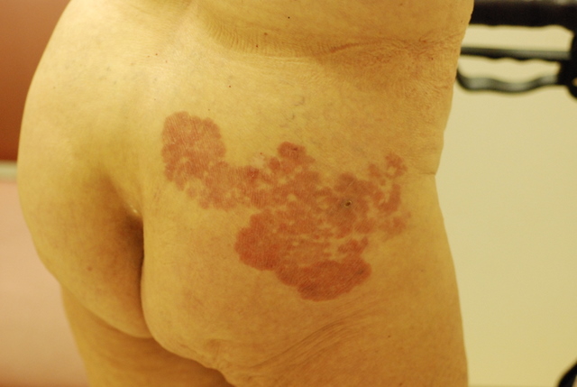
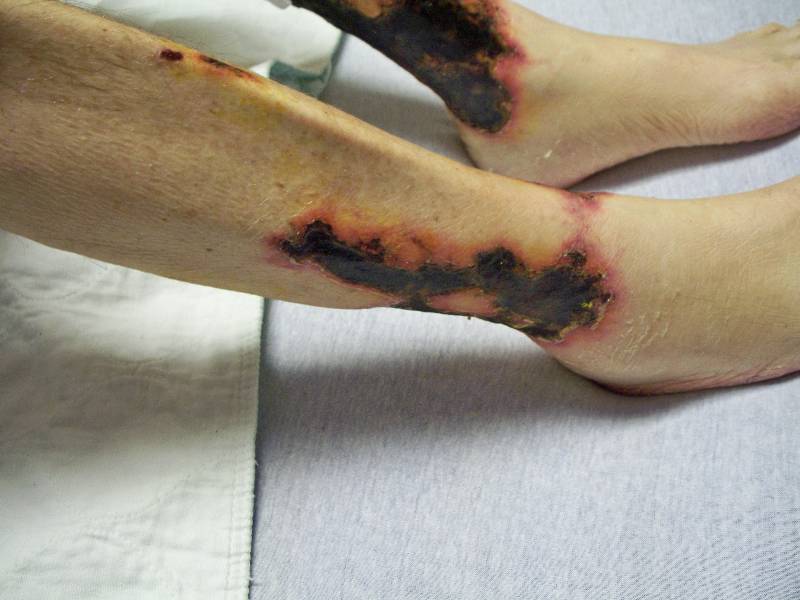
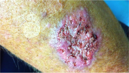
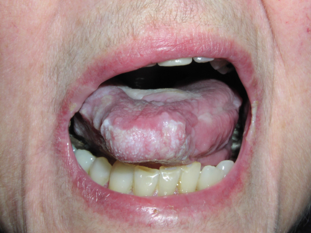
Mycosis fungoides is characterized by heterogeneous cutaneous manifestations including patches, plaques, tumors, generalized erythroderma, poikiloderma, or rarely, papules. The most common initial skin lesions are scaly patches or plaques, which can be pruritic. While most patch lesions are large (greater than 5 cm in diameter), they often vary in size, shape, and color. Lesions are often confined to the bathing trunk distribution although any body surface may be affected, even the palms and soles.
A definitive diagnosis of MF is often preceded by a “premycotic” period ranging from months to decades, during which the patient may have nonspecific, slightly scaling skin lesions and nondiagnostic biopsies. These lesions may wax and wane over the years, and a diagnosis of parapsoriasis en plaque or nonspecific dermatitis is often made.[5]
Skin biopsies demonstrate small to medium-sized atypical mononuclear cells with cerebriform nuclei infiltrating the upper dermis among epidermal keratinocytes (epidermotropism) or forming intraepidermal aggregates (Pautrier microabscesses). Pautrier microabscesses are pathognomonic, yet uncommon.[6] Spongiosis, or the collection of fluid in the epidermis, is not seen.
Immunophenotyping is used to support or confirm the results of the routine histology. The gene rearrangement tests are utilized primarily when the histology and immunophenotyping results are equivocal in patients whose clinical presentation is strongly suggestive of MF, and maybe less valuable when used to determine prognosis. [7,8]
The standard staging system for MF is based upon an evaluation of the skin (T), lymph nodes (N), visceral involvement (M), and blood. Mycosis fungoides treatment selection should be based on the disease stage. Topical, skin-directed therapies, such as topical steroids, topical retinoids, topical chemotherapy (eg, nitrogen mustard or bischloroethylnitrosourea [BCNU]), ultraviolet B (UV-B) light, UV-A light treatment enhanced with psoralen (PUVA), or electron beam radiation are indicated for early-stage disease. Systemic therapies, such as recombinant alpha-interferon, oral retinoids, monoclonal antibodies, histone deacetylase inhibitor, fusion toxin treatment, extracorporeal photopheresis) or combinations of topical and systemic therapies are indicated for patients with more advanced stages.
TREATMENT:
The patient was referred to radiology/oncology for further evaluation and treatment.
REFERENCES:
Whittaker, S. (2006). Biological insights into the pathogenesis of cutaneous T-cell lymphomas (CTCL). Seminars in Oncology, 33(1 Suppl 3), S3-S6. https://doi.org/10.1053/j.seminoncol.2006.01.004
Hall, W. W., Liu, C. R., Schneewind, O., Takahashi, H., Kaplan, M. H., Roupe, G., & Vahlne, A. (1991). Deleted HTLV-I provirus in blood and cutaneous lesions of patients with mycosis fungoides. Science, 253(5017), 317-320. https://doi.org/10.1126/science.1859792
Wood, G. S., Salvekar, A., Schaffer, J., Crooks, C. F., Henghold, W., Fivenson, D. P., Kim, Y. H., & Smoller, B. R. (1996). Evidence against a role for human T-cell lymphotropic virus type I (HTLV-I) in the pathogenesis of American cutaneous T-cell lymphoma. Journal of Investigative Dermatology, 107(3), 301-307. https://doi.org/10.1111/1523-1747.ep12353639
Pimpinelli, N., Olsen, E. A., Santucci, M., Vonderheid, E., Haeffner, A. C., Stevens, S., Burg, G., Cerroni, L., Dreno, B., Glusac, E., Guitart, J., Heald, P. W., Kempf, W., Knobler, R., Lessin, S., Sander, C., Smoller, B. S., Telang, G., Whittaker, S., Iwatsuki, K., Obitz, E., Takigawa, M., Turner, M. L., & Wood, G. S. (2005). Defining early mycosis fungoides. Journal of the American Academy of Dermatology, 53(6), 1053-1063. https://doi.org/10.1016/j.jaad.2005.08.019
Hoppe, R. T., Wood, G. S., & Abel, E. A. (1990). Mycosis fungoides and the Sezary syndrome: Pathology, staging, and treatment. Current Problems in Cancer, 14(6), 293-371. https://doi.org/10.1016/S0147-0272(90)80005-3
Jaffe, E. S., Harris, N. L., Stein, H., & Vardiman, J. W. (Eds.). (2001). World Health Organization Classification of Tumours, Pathology and Genetics of Tumours of Haematopoietic and Lymphoid Tissues. Lyon: IARC Press.
Oshtory, S., Apisarnthanarax, N., Gilliam, A. C., Cooper, K. D., & Meyerson, H. J. (2007). Usefulness of flow cytometry in the diagnosis of mycosis fungoides. Journal of the American Academy of Dermatology, 57(3), 454-462. https://doi.org/10.1016/j.jaad.2006.11.013
Juarez, T., Isenhath, S. N., Polissar, N. L., Sabath, D. E., Wood, B., Hanke, D., Haycox, C. L., Wood, G. S., & Olerud, J. E. (2005). Analysis of T-cell receptor gene rearrangement for predicting clinical outcome in patients with cutaneous T-cell lymphoma: A comparison of Southern blot and polymerase chain reaction methods. Archives of Dermatology, 141(9), 1107-1113. https://doi.org/10.1001/archderm.141.9.1107


