CORRECT DIAGNOSIS:
Diffuse Large B Cell Lymphoma
DISCUSSION:
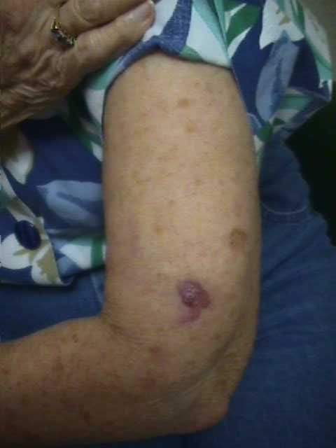
Primary cutaneous diffuse large B-cell lymphoma is an uncommon form of cutaneous lymphoma, representing 20-25% of these lymphomas, and generally having a poor prognosis.8,9 PCDLBCL are characterized by neoplastic proliferation of large, atypical B-lymphocytes, which clinically are most commonly present on the lower legs and are described as rapidly growing red-brown plaques.10 Typically, at the time of diagnosis, PCDLBCL has no extracutaneous involvement and is confined only to the skin.11 Nevertheless, PCDLBCL has a propensity to spread to lymph nodes and extracutaneous sites.12
Cutaneous diffuse large B-cell lymphoma has the histological characterization of a diffuse infiltrate throughout the entire dermis and extending into the subcutaneous, usually sparing a thin subepidermal grenz zone, and consisting of large B-cells, which were formally known as centroblasts and immunoblasts.12 Immunohistochemical stains of the neoplastic cells in PCDLBCL are typically positive for CD19, CD20, CD79a, Bcl-2, and MUM-1.13 There is an excellent correlation between CD20 and PAX-5 expression. PAX-5 is a B-cell specific activator protein (BSAP). PAX-5 is expressed primarily in pro-, pre-, and mature B-cells, but not in plasma cells. Anti-PAX-5 has the ability to detect all committed B-cells. It is very specific to B-cell lineage and does not stain T-cells.
The characteristic predominance of the large tumor cells leads to the aggressiveness of this subtype. The histologic predominance of large cells with round nuclei as opposed to large cells with cleaved nuclei have a less favorable prognosis.14 Additionally, the expression of Bcl-2 protein also has been identified as an adverse prognostic factor.15 More recently, one study showed that the 5-year survival rate was decreased to a predicted 27% with the deletion of chromosome region 9p21, compared to 100% in cases without deletion.16 However, the location on the leg of PCDLBCL is the main negative prognostic factor for cutaneous lymphomas, representing a 3-year survival rate of 43% compared to 77% in non-leg anatomic locations.4 Furthermore, non-leg PCDLBCL carries a less favorable prognosis than primary cutaneous marginal zone B-cell lymphoma and primary cutaneous follicle center lymphomas, and should, therefore, be grouped in classification with primary cutaneous large B cell lymphoma leg type, as proposed by the WHO-EORTC classification.4,11
Radiotherapy is considered the treatment of choice in the management of primary cutaneous lymphomas, with 2 cm margins.2 There has also been reported success with the use of anthracycline-containing chemotherapies and rituximab, a chimeric monoclonal IgG1 antibody that is directed against the CD20 antigen of B cells.17,18 Additionally, rituximab has been shown to be effective in overcoming Bcl-2-associated resistance to chemotherapy, which is a poor prognostic factor when expressed.19,20
TREATMENT:
The tumor was surgically removed by surgical oncology and received external beam radiation and chemotherapy. The patient was worked up by oncology for evaluation of any internal malignancy as well, which was found to be negative. In the subsequent years, she further developed diffuse large B-cell lymphoma of the left lower leg and left arm, demonstrating a long history of left upper extremity B-cell lymphoma which required multiple courses of external beam radiation and chemotherapy.
CONCLUSION:
Primary cutaneous diffuse large B-cell lymphomas (PCDLBCL) are defined as malignant atypical B-cell proliferations presenting with cutaneous involvement alone and no evidence of extracutaneous manifestations at time of diagnosis. It most commonly presents in elderly women and are often located on the lower legs. We presented a case of an 80-year-old Caucasian female who noticed a nodular lesion on her left arm and in subsequent years left lower leg that rapidly enlarged in a few months. Histological and immunophenotypical features were compatible with PCDLBCL. We also highlighted the new classification system of primary cutaneous B-cell lymphomas as well as provided a brief overview on prognosis and management.
REFERENCES:
Newton, R., Ferlay, J., Beral, V., & Devesa, S. S. (1997). The epidemiology of non-Hodgkin’s lymphoma: Comparison of nodal and extra-nodal sites. International Journal of Cancer, 72(6), 923-930. https://doi.org/10.1002/(SICI)1097-0215(19970307)72:6<923::AID-IJC24>3.0.CO;2-Q [PMID: 9130083]
Garbea, A., Dippel, E., Hildenbrand, R., et al. (2002). Cutaneous large B-cell lymphoma of the leg masquerading as chronic venous ulcer. British Journal of Dermatology, 146, 144-147. https://doi.org/10.1046/j.1365-2133.2001.04637.x [PMID: 11872190]
Hembury, T. A., Lee, B., Gascoyne, R. D., et al. (2002). Primary cutaneous diffuse large B-cell lymphoma: A clinicopathologic study of 15 cases. American Journal of Clinical Pathology, 117, 574-580. https://doi.org/10.1309/2G4Y-0R5V-GG5X-PQEA [PMID: 11916806]
Grange, F., Beylot-Barry, M., Courville, P., et al. (2007). Primary cutaneous diffuse large B-cell lymphoma, leg type. Archives of Dermatology, 143(9), 1144-1150. https://doi.org/10.1001/archderm.143.9.1144 [PMID: 17846276]
Golling, P., Cozzio, A., Dummer, R., et al. (2008). Primary cutaneous B-cell lymphomas: Clinicopathological, prognostic, and therapeutic characterization of 54 cases according to the WHO-EORTC classification and the ISCL/EORTC TNM classification system for primary cutaneous lymphomas other than mycosis fungoides and Sézary syndrome. Leukemia & Lymphoma, 49(6), 1094-1103. https://doi.org/10.1080/10428190801917638 [PMID: 18421173]
Connors, J. M., HIS, E. D., & Foss, F. M. (2002). Lymphoma of the skin. In Hematology (pp. 263-282).
Moore, M. M., Kovich, O. I., & Brown, L. H. (2007). Primary cutaneous B-cell lymphoma (low grade, non-large cell). Dermatology Online Journal, 13(1), 8. https://doi.org/10.5070/D13s1 [PMID: 17383155]
Senff, N. J., Noordijk, E. M., Kim, Y. H., et al. (2008). European Organization for Research and Treatment of Cancer and International Society for Cutaneous Lymphoma consensus recommendations for the management of cutaneous B-cell lymphomas. Blood, 112(5), 1600-1609. https://doi.org/10.1182/blood-2007-12-122520 [PMID: 18578076]
Zinzani, P. L., Quaglino, P., Pimpinelli, N., et al. (2006). Prognostic factors in primary cutaneous B-cell lymphoma: The Italian Study Group for Cutaneous Lymphomas. Journal of Clinical Oncology, 24(7), 1376-1382. https://doi.org/10.1200/JCO.2005.02.2197 [PMID: 16525102]
Cerroni, L., Gatter, H., & Kerl, H. (2005). Large B-cell lymphoma, leg type. In L. Cerroni, K. Gatter, & H. Kerl (Eds.), An illustrated guide to skin lymphoma (pp. 112-116). Oxford: Blackwell Publishing.
Willemze, R., Jaffe, E. S., Burg, G., et al. (2005). WHO-EORTC classification of cutaneous lymphomas. Blood, 106(7), 2491-2497. https://doi.org/10.1182/blood-2005-02-0466 [PMID: 16000316]
Kodama, K., Massone, C., Chott, A., et al. (2005). Primary cutaneous large B-cell lymphomas: Clinicopathologic features, classification, and prognostic factors in a large series of patients. Blood, 106(7), 2491-2497. https://doi.org/10.1182/blood-2005-03-1403 [PMID: 16000316]
Paulli, M., Viglio, A., Vivenza, D., et al. (2002). Primary cutaneous large B-cell lymphoma of the leg: Histogenetic analysis of a controversial clinicopathologic entity. Human Pathology, 33(9), 937-943. https://doi.org/10.1016/S0046-8177(02)80417-3 [PMID: 12270670]
Grange, F., Bekkenk, M. W., Wechsler, J., et al. (2001). Prognostic factors in primary cutaneous large B-cell lymphomas: A European multicenter study. Journal of Clinical Oncology, 19(16), 3602-3610. https://doi.org/10.1200/JCO.2001.19.16.3602 [PMID: 11579173]
Grange, F., Petrella, T., Beylot-Berry, M., et al. (2004). Bcl-2 protein expression is the strongest independent prognostic factor of survival in primary cutaneous large B-cell lymphomas. Blood, 103(10), 3662-3668. https://doi.org/10.1182/blood-2003-06-1938 [PMID: 14630539]
Senff, N. J., Zoutman, W. H., Vermeer, M. H., et al. (2007). Fine mapping chromosomal loss at 9p21: Correlation with prognosis in primary cutaneous large B-cell lymphoma, leg type. Journal of Investigative Dermatology, 127(Suppl), S92. https://doi.org/10.1038/sj.jid.5700675 [PMID: 17646878]
Grange, F., Maubec, E., Bagot, M., et al. (2009). Treatment of cutaneous B-cell lymphoma, leg type, with age-adapted combinations of chemotherapies and rituximab. Archives of Dermatology, 145(3), 329-330. https://doi.org/10.1001/archderm.145.3.329 [PMID: 19204129]
Garcia, C. P., Florez, A., Pardavilla, R., et al. (2009). Primary cutaneous large B-cell lymphoma, leg type, successfully treated with rituximab plus chemotherapy. European Journal of Dermatology, 19(4), 394-395. https://doi.org/10.1684/ejd.2009.0635 [PMID: 19746376]
Coiffier, B., Lepage, E., Brière, J., et al. (2002). CHOP chemotherapy plus rituximab compared with CHOP alone in elderly patients with diffuse large B-cell lymphoma. New England Journal of Medicine, 346(4), 235-242. https://doi.org/10.1056/NEJMoa011792 [PMID: 11784887]
Mounier, N., Brière, J., Gisselbrecht, C., et al. (2003). Rituximab plus CHOP (R-CHOP) overcomes bcl-2 associated resistance to chemotherapy in elderly patients with diffuse large B-cell lymphoma. Blood, 101(11), 4279-4284. https://doi.org/10.1182/blood-2002-07-2286 [PMID: 12667971]


