CORRECT DIAGNOSIS:
Nevus lipomatosus superficialis
DISCUSSION:
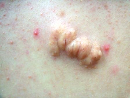
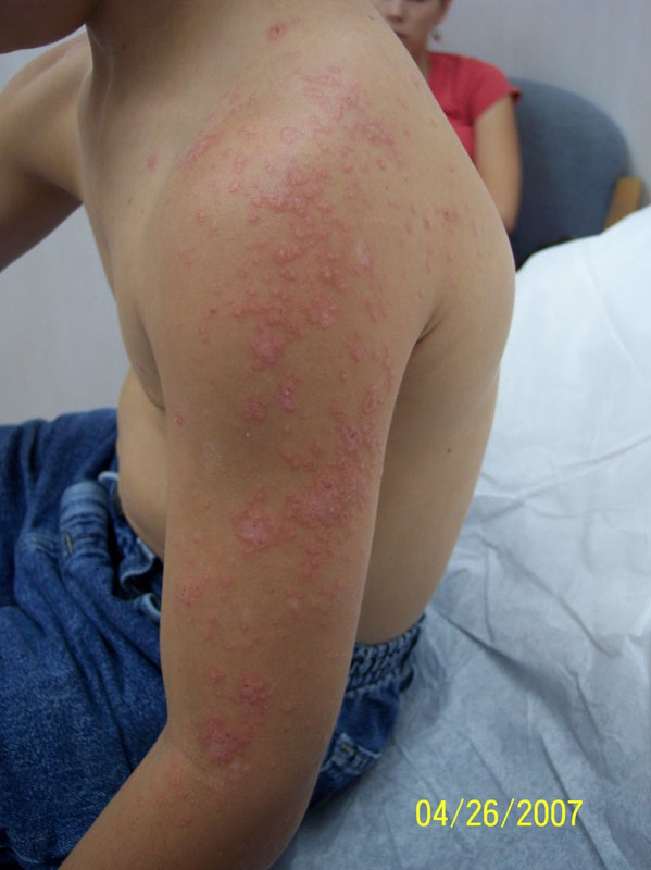
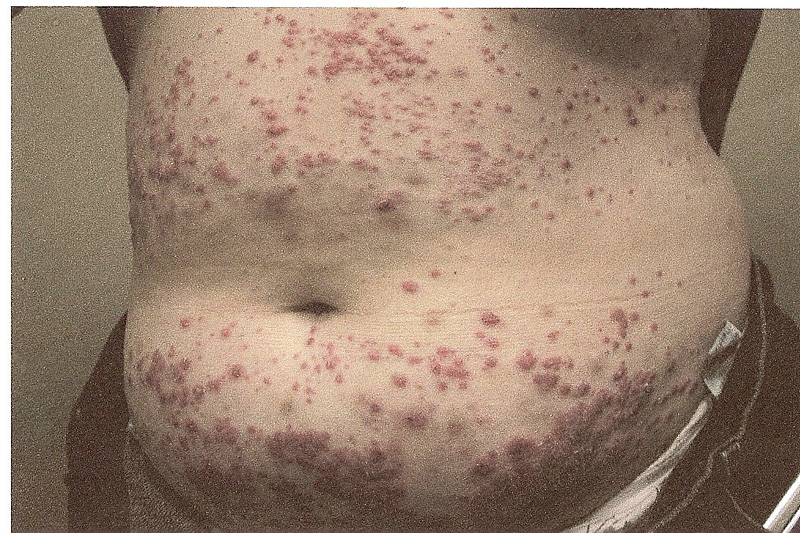
Nevus lipomatosus superficialis (NLCS), also known as nevus lipomatosus of Hoffmann-Zurhelle, was first described in 1921.(1) This lesion presents at birth or early childhood as a flesh-colored or yellow papule with a smooth or corrugated surface. This lesion favors the pelvic girdle but has been reported in a wide variety of anatomical locations. These lesions are not usually associated with other developmental abnormalities. (2)
Two clinical variations of NLCS have been described in the literature. The first is the classic or multiple types. This appears from birth to over the next two decades of life and is a flesh-colored to the yellow plaque with a smooth or corrugated surface following natural cleavage lines of the skin. The classic usually occurs on the sacral, lumbar, or pelvic girdle regions.(1-3) The second is the solitary type, which may not appear until the fifth decade of life.(4) They present as small solitary nodules similar to skin tags appear over the arms, knees, axillae, ears, and scalp.(1)
The pathogenesis of NLCS is unknown but there are several theories. These theories include; adipose metaplasia during the course of degenerative changes in dermal connective tissue, developmental displacement of adipocytes, developmental displacement of mature adipocytes from the dermal vessel walls.(1,2)
Histopathological diagnosis is made by the presence of ectopic mature adipocytes in the dermis. Varying amounts of adipose tissue may be present and routinely does not connect with the underlying subcutaneous fat. Fat can comprise anywhere from 10-70% of the lesion. Thickening of the collagen bundles increased deeper elastic tissue and decreased superficial elastic tissue, and increased blood vessels in the papillary dermis may also be present.(5)
TREATMENT:
The treatment of choice is surgical excision, but treatment is not necessary.
REFERENCES:
Khandpur, S., Nagpal, S., Chandra, S., et al. (2009). Giant nevus lipomatosus cutaneous superficialis. Indian Journal of Dermatology, Venereology, and Leprology, 74(4), 407-408. https://doi.org/10.4103/0378-6323.57858 [PMID: 19734338]
Lane, J. E., Clark, E., & Marzec, T. (2003). Nevus lipomatosus cutaneus superficialis. Pediatric Dermatology, 20(4), 313-314. https://doi.org/10.1046/j.1525-1470.2003.20303.x [PMID: 14506415]
Park, M., & Kim, Y. (2009). A soft lesion on the scrotum: A quiz. Acta Dermato-Venereologica, 89(5), 549-550. https://doi.org/10.2340/00015555-0665 [PMID: 19772015]
Ragsdale, B. (2005). Tumors with fatty, muscular, osseous, and cartilaginous differentiation. In D. E. Elder, R. E. Leuitsas, B. L. Johnson Jr., & G. F. Murphy (Eds.), Lever’s histopathology of the skin (pp. 1061-1107). Philadelphia, PA: Lippincott Williams and Wilkins.
Weedon, D. (2010). Tumors of fat. In Weedon skin pathology (pp. 846). Elsevier Limited.


