CORRECT DIAGNOSIS:
Hidradenitis Suppurativa
DISCUSSION:
Hidradenitis suppurativa was first named by Verneuil in 1854. Initially, apocrine gland dysfunction was considered to be the underlying cause; we now know that occlusion of the follicular infundibula followed by rupture of the follicle is the inciting event. Rupture of the follicle allows the introduction of its contents, including keratin and bacteria, into the surrounding dermis. This excites a vigorous chemotactic response and abscess formation. Epithelial strands are generated, possibly from ruptured follicular epithelia, and form sinus tracts.
Hidradenitis suppurativa starts at or soon after puberty. Women are affected three times as often as men. People of African descent have a higher incidence than those of European descent. Despite the apparent strong clinical influence of sex hormones, most patients have normal androgen profiles. In addition, apocrine glands (unlike sebaceous glands) are not androgen-sensitive. Although a familial form of hidradenitis suppurativa with autosomal dominant inheritance has been described, studies have found no significant associations with specific human leukocyte antigen (HLA)-A, -B or -DR subtypes. Lithium therapy has been reported to exacerbate hidradenitis suppurativa.
Initially, inflammatory nodules and sterile abscesses develop in the axillae, groin, perianal and/or inframammary areas. These lesions may be very tender and extremely painful. Over time, sinus tracts and hypertrophic scars may develop. This is accompanied by chronic drainage which can be malodorous. The discharged fluid is often a mixture of serous exudate, blood and pus, in varying proportions. Complications of hidradenitis suppurativa include anemia, secondary amyloidosis, lymphedema, and fistulae to the urethra, bladder, peritoneum, and rectum. Other complications include hypoproteinemia, nephrotic syndrome, and arthropathy. Squamous cell carcinoma, sometimes with metastasis, maybe an occasional complication of chronic scarring disease.
There is a heavy mixed inflammatory cell infiltrate in the lower half of the dermis, usually with extension into the subcutis. Abscesses are present in active cases, and they may connect to the skin surface via a sinus tract. The sinus tracts contain inflammatory cells and keratinous debris. Granulation tissue with inflammatory cells and occasional foreign body giant cells is present in up to 25% of biopsy specimens. In chronic disease, there may be extensive fibrosis with the destruction of pilosebaceous follicles and sweat glands. Staphylococcal furunculosis, Crohn’s disease, granuloma inguinale, mycetoma, tuberculosis, and elephantiasis nostras verrucosa may resemble hidradenitis suppurativa.
Medical treatment includes weight reduction (if the patient is overweight) and measures to reduce friction and moisture (loose undergarments, absorbent powder, antiseptic soaps, and topical aluminum chloride). Triamcinolone (5 mg/ml) may be injected intralesionally into early inflammatory lesions. The chronic use of topical clindamycin has proven beneficial in some patients and may reduce Staphylococcus aureus carriage and secondary infection; 5-day courses of intranasal mupirocin are used in nasal carriers of S. aureus. Incision and drainage should be minimized because it may result in scarring and chronic sinus tract formation. Systemic corticosteroids (prednisone 60-80 mg/day) often lead to dramatic improvement, but the condition usually flares when the medication is discontinued. Cyproterone acetate has been effective in some cases, but high doses were necessary, which raises safety concerns. A combination of cyproterone acetate plus ethinyl estradiol (30ìg/day for 21 days) worked better than either drug alone. Isotretinoin has not been particularly effective, but acitretin (25 mg twice daily) has anecdotally been reported as beneficial. Finasteride (5 mg/day for 3 months) has led to an improvement in some patients.
The proinflammatory cytokine, TNF-á, may be involved in the pathogenesis of hidradenitis. This has led to numerous case reports of the use of TNF-á inhibitors as a possible treatment. These reports have yielded mixed results. Most patients improved in the short-term, but most relapsed later. In both of these studies, adverse drug reactions were seen in a significant minority of patients. The data appear to suggest that TNF-á inhibitors, especially infliximab, may have some short-term benefit for treating hidradenitis, but maintenance infusions may be necessary to maintain improvement. The best results seem to occur when the involved areas are excised and closed primarily or grafted. The earlier in the course of the disease that this is performed, the better. Surgical treatment of chronic hidradenitis suppurativa with CO2 laser stripping and secondary intention healing has proven to be quite effective. However, the wounds may require months to heal and the patient needs to be fully informed.
Other agents used in the treatment of hidradenitis suppurativa include cyclosporine as well as botulinum toxin A (to reduce sweating). Successful outcomes are based upon case reports and small series of patients.
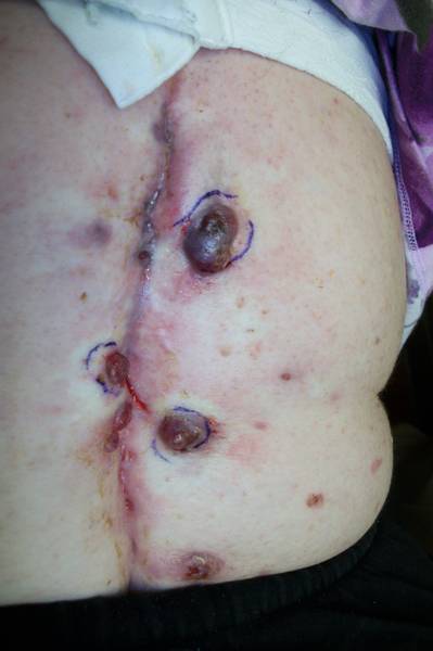
This case is presented for the atypical presentation. The literature reports a little over 10 cases of atypical presentations of hidradenitis suppurativa presenting on the back and/or face.
TREATMENT:
The patient received intralesional corticosteroids at the time of biopsy. The case was discussed with local experts and we were planning on starting an anti-TNF agent. Unfortunately, the patient did not return for follow up.
REFERENCES:
McMichael, A., Sanchez, D. S., & Kelly, P. (2011). Folliculitis and the follicular occlusion tetrad. In J. L. Bolognia, J. L. Jorizzo, & R. P. Rapini (Eds.), Dermatology (2nd ed.). Elsevier Inc.
Attanoos, R. L., Appleton, M. A., & Douglas-Jones, A. G. (1995). The pathogenesis of hidradenitis suppurativa: A closer look at apocrine and apoeccrine glands. British Journal of Dermatology, 133(2), 254-258. doi:10.1111/j.1365-2133.1995.tb04616.x
Poli, F., Wolkenstein, P., & Revuz, J. (2010). Back and face involvement in hidradenitis suppurativa. Dermatology, 221(2), 137-141. doi:10.1159/000313237
Jemec, G. B., & Hansen, U. (1996). Histology of hidradenitis suppurativa. Journal of the American Academy of Dermatology, 34(6), 994-999. doi:10.1016/S0190-9622(96)90018-2


