CORRECT DIAGNOSIS:
Basal Cell Nevus Syndrome (Gorlin Syndrome)
DISCUSSION:
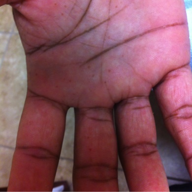
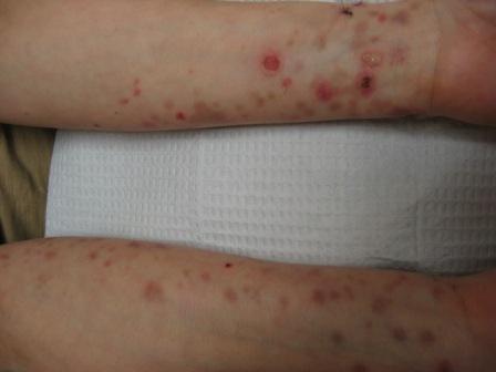
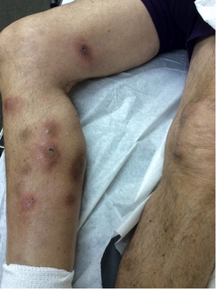
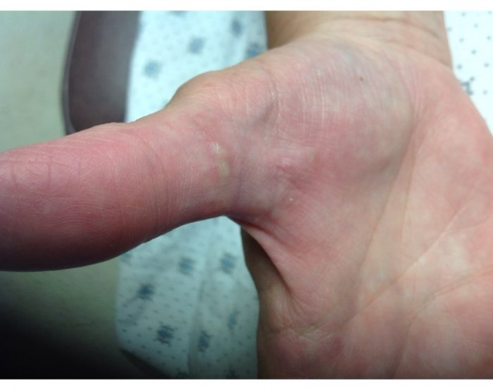
Basal cell nevus syndrome (BCNS) was first reported in 1894 but the clinical manifestations were more clearly defined in 1960 by Gorlin and Goltz (1). It is an autosomal dominant disorder with malignant potential that is caused by mutations in the PTCH1 gene (2). Normally, PTCH is bound to smoothened and inhibits nuclear transcription and cellular proliferation (2). With inactivating mutations of PTCH1, smoothened is no longer inhibited thus causing increased cellular proliferation (2). Vismodegib is an inhibitor of smoothened thus preventing nuclear transcription (2). Clinically, patients may present with multiple developmental anomalies such as frontal bossing and skeletal abnormalities during birth and jaw cysts along with palmoplantar pits by childhood (2). Of all BCNS case reports, only 5% involve African Americans (3). In BCNS, BCCs may resemble angiomas, skin tags, or melanocytic nevi (2). They can appear on any part of the body but occur more frequently on sun-exposed sites (4). Extracutaneous manifestations can include ectopic calcification of the falx cerebri, agenesis of the corpus callosum, medulloblastomas and meningiomas, as well as mental retardation (5). Genitourinary features include ovarian fibromas and fibrosarcomas (5). Patients also may present with hypertelorism, congenital blindness, cataracts, colobomas, and strabismus (5). Abnormalities in the musculoskeletal system include macrocephaly, frontal bossing, bifid ribs, vertebral fusion, and kyphoscoliosis (5). The lifespan of a patient with BCNS is normal if the BCCs are treated early and if no other malignancies develop (4). Yearly panoramic radiographs of the jaw are recommended with complete removal of identified odontogenic keratocysts as they can be extremely aggressive and even undergo malignant transformation (2). Radiologic evaluation should be performed to check for calcification of the falx cerebri, as well as abnormalities of the ribs, vertebrae, and phalanges (2). An MRI of the entire neurological axis may be warrented due to the wide range of skeletal anomalies present in BCNS (2). Additional imaging modalities to consider include transvaginal ultrasound for pelvic masses and MRI of the brain (2).
In conclusion, BCNS is a fairly common genodermatosis, but only rarely occurs in African Americans. This case illustrates the importance of evaluating patients thoroughly who may present with “unusual” locations of basal cell cancers. Vismodegib may plan an important role in decreasing the tumor burden and shrinking the size of the tumors, but a high rate of side effects will likely prevent its long-term use in patients with lifelong BCNS.
TREATMENT:
Therapy is directed at the individual lesions as they arise, and the best treatment modality depends on clinical presentation, cell type, tumor size, and location (4). Treatment options include surgical excision, ED&C, cryotherapy, topical 5-fluorouracil, or imiquimod (4). Oral retinoids can be given to suppress new BCCs (4). Patients should be counseled about daily use of broad-spectrum sunblock and strict sun avoidance (4). Vismodegib, an inhibitor of smoothened, is an FDA-approved oral medication for the treatment of locally advanced or metastatic BCCs. Vismodegib appears to be an excellent option for patients with BCNS in whom multiple surgeries for hundreds of tumors result in disfiguring scars. In a recent original article from the NEJM, it was indeed found that inhibiting the smoothened pathway with Vismodegib in patients with BCNS decreased the incidence of new BCCs that were eligible for surgical excision (6). Some patients even had complete regression of all BCCs as well as the disappearance of palmar and plantar pits (6). Side effects caused 54% of patients to prematurely stop the medication and included muscle spasms, alopecia, dysgeusia, and weight loss (6, 7). Thus, long-term treatment of patients with BCNS with Vismodegib does not seem feasible owing to the large drop-out rate caused by side effects.
REFERENCES:
Gorlin, R. J., & Goltz, R. W. (1960). Multiple nevoid basal-cell epithelioma, jaw cysts and bifid rib. A syndrome. N Engl J Med, 262, 908-912. PMID: 13820268
Bolognia, J. L., Jorizzo, J. L., & Schaffer, J. V. (2012). Dermatology (3rd ed.). Elsevier Saunders: 813, 1151, 1776-1777, Table 108.5.
Goldstein, A. M., Pastakia, B., DiGiovanna, J. J., et al. (1994). Clinical findings in two African-American families with the nevoid basal cell carcinoma syndrome (NBCC). Am J Med Genet, 50, 272. PMID: 8029176
Habif, T. P. (2010). Clinical Dermatology: A Color Guide to Diagnosis and Therapy (5th ed.). Mosby Elsevier: 808-811.
Spitz, J. L. (2005). Genodermatoses: A Clinical Guide to Genetic Skin Disorders (2nd ed.). Philadelphia, PA: Lippincott Williams & Wilkins: 170-173.
Tang, J. Y., Mackay-Wiggan, J. M., Aszterbaum, M., et al. (2012). Inhibiting the Hedgehog Pathway in Patients with the Basal-Cell Nevus Syndrome. N Engl J Med, 366, 2180-2188. PMID: 22716924
Sekulic, A., Migden, M. R., et al. (2012). Efficacy and Safety of Vismodegib in Advanced Basal Cell Carcinoma. N Engl J Med, 366, 2171-2179. PMID: 22716923


