CORRECT DIAGNOSIS:
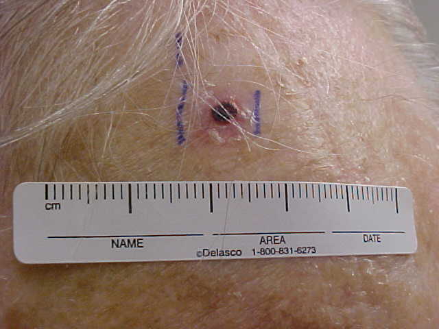
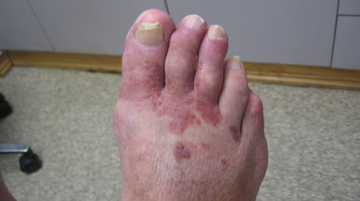
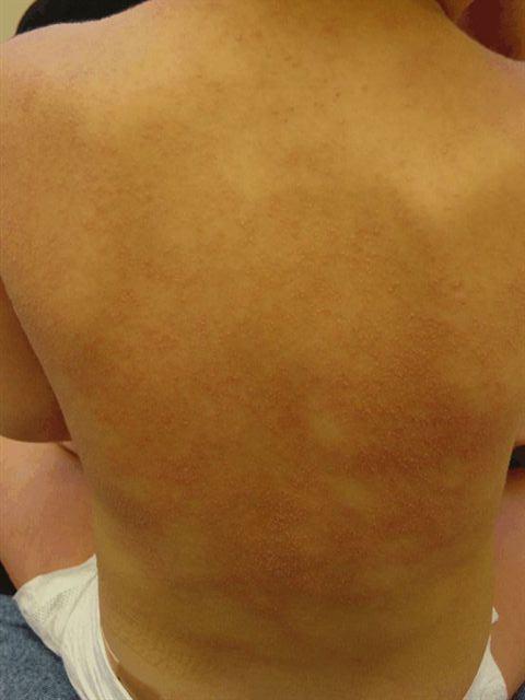
Eruptive Keratoacanthomas, in regression, arising in a tattoo
DISCUSSION:
Keratoacanthomas are rapidly-growing epithelial tumors that arise in hair follicles, most frequently after trauma to sun-damaged skin. There is disagreement about whether these lesions should be considered benign or a malignant variant of squamous cell carcinoma. It has been suggested that keratoacanthomas are the benign self-resolving counterpart to the malignant so-called infundibulocystic squamous cell carcinoma. Whether these lesions are reactive or neoplastic is also not entirely clear. Keratoacanthomas have shown a wide range of clinical behaviors from spontaneous regression to local aggression with malignant transformation and rarely metastasis.
Multiple predisposing risk factors have been associated with the development of keratoacanthomas, including trauma, immunosuppression (organ transplantation, chemotherapy), chronic ultraviolet light exposure, Fitzpatrick skin types I and II, human papillomavirus infection, and genetic predisposition (Muir-Torre syndrome, xeroderma pigmentosum). Trauma-induced lesions have been reported within thermal burns, friction burns, vaccination sites, emanating from razor cuts, laser resurfacing, skin grafts, and arthropod assaults. They typically develop between 1 week and 1-year post-trauma.
In our case, the trauma from the tattoo was the likely inciting factor. However, sensitization from prior tattoos, chronic sun exposure due to living in South Florida, and occupational exposure to carcinogenic agents at the World Trade Center may have played a contributing role. The diagnosis was challenging because the clinical differential diagnosis also included viral wart inoculation and koebnerization, squamous cell carcinomas, granulomas, and infection with mycobacacteria or fungus. Dermatoscopic evaluation was difficult due to the presence of tattoo pigment.
In 1955, Ereaux et al reported on the first case of multiple keratoacanthomas preceded by trauma in a patient with a gasoline burn on his extremities. In 1966, McQuarrie reported a squamous cell carcinoma arising in a tattoo. Since then, keratin-producing tumors in association with tattoos have been well described in the literature with findings that overlap with ours, although they remain an uncommon phenomenon when compared with other tattoo reactions.
Interstingly, our patient’s keratoacanthomas were restricted to the sites of red pigment, as has been most commonly reported in the literature. The composition of ink may play a crucial role in the pathologic process. The pigments commonly employed in red ink are mercuric sulfide (cinnabar and vermilion), scarlet lake (organic pigment), carmine (dried insect bodies), cochinilla, cadmium red (selenide), and azo dyes. After World War II, red tattoo ink contained high quantities of mercury to which many reactions have been attributed. In 1976, after calls for increased regulation, the FDA limited the amount of mercury in tattoo pigments to 3 parts per million. Various other pigments are often now combined to create different red hues, but red inks still cause more reactions than other colors. In a recent case series of eleven keratoacanthomas, red tattoo ink was indeed the most common, occurring in 82% of cases, although reactions to all colors have been reported. Since the FDA has not approved any ink for tattooing, the precise composition of tattoo pigments is not regulated and difficult to analyze. Additionally, red is one of the most commonly used colors in tattoos and the high prevalence may also reflect the frequency of its use.
While the malignancy of keratoacanthomas is a matter of debate, a recent review uncovered 50 cases of malignant melanoma associated with tattoos. Although this number is low enough to be considered coincidental, there has been an increase in reported cases. As popularity of tattooing grows and composition of ink continues to change and evolve, coincidental malignant lesions will become more likely and new inks with as-yet-unknown carcinogenic potential raise the importance for dermatologists to be familiar with tattoo ink toxicology as it relates to skin tumors, sun habits, the challenges of skin cancer screenings, and other relevant health issues.
TREATMENT:
Various topical, systemic, and surgical approaches have been used in the treatment of keratoacanthomas. These include topical and systemic retinoids, intralesional bleomycin and 5-fluorouracil, methotrexate, cyclophosphamide, curettage, cryosurgery, laser ablation, radiation therapy, and surgical excision.
With the biopsy pending, we proceeded with two 15-second cycles of liquid nitrogen cryosurgery to each lesion. Over the course of four monthly treatments, there was interval improvement with regression of all lesions and minimal dyspigmentation or scarring. Half of the lesions regressed completely without recurrences, and all others have diminished in size.
Some authors argue that surgical removal of the entire hyperproliferative area is necessary and should be followed with a histological examination to exclude the possibility of a squamous cell carcinoma. Indeed, verrucous carcinoma has been shown to develop at the site of inflammatory or scarring processes and it may be difficult to distinguish in certain cases from keratoacanthoma. However, in our case, with multiple lesions contained within a small surface area, aesthetic concerns, and a favorable response to empiric cryosurgery, surgical treatment was not a favored option. Instead, we recommended continued monitoring in order to manage high risk lesions should any develop.
REFERENCES:
Balfour, E., Olhoffer, I., Leffell, D., & Handerson, T. (2003). Massive pseudoepitheliomatous hyperplasia: An unusual reaction to a tattoo. American Journal of Dermatopathology, 25, 338–340. PMID: 12815052
Chorny, A., Stephens, F., & Cohen, J. (2007). Eruptive keratoacanthomas in a new tattoo. Archives of Dermatology, 143, 1457. PMID: 17998473
Dessoukey, M. W., Omar, M. F., & Abdel-Dayem, H. (1997). Eruptive keratoacanthomas associated with immunosuppressive therapy in a patient with systemic lupus erythematosus. Journal of the American Academy of Dermatology, 37, 478–480. PMID: 9263065
Ereaux, L. P., Schopflocher, P., & Fournier, C. J. (1955). Keratoacanthoma. Archives of Dermatology, 71, 73–83. PMID: 13263381
Gewirtzman, A., Meirson, D. H., & Rabinovitz, H. (1999). Eruptive keratoacanthomas following carbon dioxide laser resurfacing. Dermatologic Surgery, 25, 666–668. PMID: 10422254
Ghadially, F. N. (1961). The role of the hair follicle in the origin and evolution of some cutaneous neoplasms of man and experimental animals. Cancer, 14, 801–816. PMID: 13737400
Grine, R. C., Hendrix, J. D., & Greer, K. E. (1997). Generalized eruptive keratoacanthoma of Grzybowski: Response to cyclophosphamide. Journal of the American Academy of Dermatology, 36, 786–787. PMID: 9118208
Hirsch, R. J., Wall, T. L., Avram, M. M., & Anderson, R. R. (2008). In Dermatology (2nd ed., J. L. Bolognia, J. L. Jorizzo, & R. P. Rapini, Eds.). Mosby/Elsevier.
Karaa, A., & Khachemoune, A. (2007). Keratoacanthoma: A tumor in search of a classification. International Journal of Dermatology, 46, 671–678. PMID: 17999380
Kossard, S., Kong-Bing, T., & Choy, C. (2008). Keratoacanthoma and infundibulocystic squamous cell carcinoma. American Journal of Dermatopathology, 30, 127. PMID: 18356840
McQuarrie, D. G. (1966). An unusual breast cancer: Squamous-cell carcinoma arising in a tattoo. Minnesota Medicine, 49, 799.
Mortimer, N. J., Chave, T. A., & Johnston, G. A. (2003). Red tattoo reactions. Clinical and Experimental Dermatology, 28, 508. PMID: 14617252
Paradisi, A., Capizzi, R., De Simone, C., Fossati, B., Proietti, I., & Amerio, P. L. (2006). Malignant melanoma in a tattoo: Case report and review of the literature. Melanoma Research, 16, 375–376. PMID: 16715003
Pattee, S. F., & Silvis, N. G. (2003). Keratoacanthoma developing in sites of previous trauma: A report of two cases and review of the literature. Journal of the American Academy of Dermatology, 48, S35. PMID: 12686571
Schwartz, R. A. (1995). Verrucous carcinoma of the skin and mucosa. Journal of the American Academy of Dermatology, 32, 1. PMID: 7799208
Sowden, J. M., Byrne, J. P. H., Smith, A. G., et al. (1991). Red tattoo reactions: X-ray microanalysis and patch-test studies. British Journal of Dermatology, 124, 576. PMID: 2011944
Vitiello, M., Echeverria, B., Romanelli, P., Abuchar, A., & Kerdel, F. (2010). Multiple eruptive keratoacanthomas arising in a tattoo. Journal of Clinical and Aesthetic Dermatology, 3, 54–55.
Washington, C. V., & Mikhail, G. R. (1987). Eruptive keratoacanthoma en plaque in an immunosuppressed patient. Journal of Dermatologic Surgery and Oncology, 13, 1357–1360. PMID: 2821873


