CORRECT DIAGNOSIS:
Basal Cell Carcinoma
DISCUSSION:
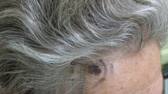
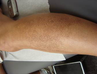
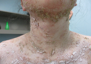
We report the case of a 68-year-old man who had multiple small soft, skin-colored round and oval pedunculated papillomas with peri-lesional erythema at both the axillae and around his neck. Clinically, a diagnosis of multiple acrochordons was made. A shave biopsy was performed per the patient’s request. Microscopic examination of the specimen contained discrete nodular masses of basaloid cells arising at several different sites from the basal cell layer. The tumor demonstrated other features characteristic of basal cell carcinoma, such as uniform cellularity, peripheral palisading, and a distinctive connective tissue stroma. The patient underwent Mohs surgery with complete removal of the lesion.
Skin tags (also known as acrochordon, cutaneous tag, papilloma colli, fibroma pendulum, cutaneous papilloma, fibroma molluscum, templeton skin tags) is among the most common benign skin lesions. It has a reported incidence of 46 percent in the general population, although it seems likely that the “life –time prevalence” of this lesion may reach 100 percent. It is more prevalence in overweight or obese patient. Interestingly, the presence of skin tags may be an important marker for the presence of type II diabetes mellitus(4). These skin tags are often seen frequently in the neck, axillae and on the eyelids, and less often on the trunk and groin (5). They are small, flesh-colored to dark brown, pinhead-sized and larger, sessile and pedunculated papillomas (5). Histologically, a skin tag shows localized hyperplasia of the dermis with loosely arranged collagen fibers and dilated capillaries and lymphatic vessels (7). Patients are motivated to remove skin tags due to cosmetic appearance but the health care infrastructure is often less interested because most of them are benign and the cost of the clinical intervention can be hard to justify. This is usually accomplished by simple snipping with scissors, electrodessication or cryosurgery. Pathologic evaluation of the removed growth is often not done, as these lesions are almost universally assumed to be benign.
In 1996, Eads, Chuang, Fabre, Farmer and Hood concluded in their study that cutaneous lesions diagnosed as typical fibroepithelial polyps by dermatologists need not be submitted for microscopic examination(6), even though they found five malignant lesions, four basal cell carcinoma and one squamous cell carcinoma in situ, out of 1335 clinical specimens submitted as skin tags.
In contrast, acrochondron is a presenting sign of nevoid basal cell carcinoma syndrome, especially in children (5). Therefore, acrochondron in a child may be a sign leading to early diagnosis of this syndrome and deserves biopsy. In 1993, Hayes A. and Berry A. reported the first case of basal cell carcinoma arising in a fibroepithelial polyp in a 59-year-old woman. In one recent report by Schwart R, Tarlow M and Lambert C on karatoacathoma-like squamous cell carcinoma within the fibroepithelial polyp in a 53-year-old white man.
This patient and previous cases illustrates that both squamous cell carcinoma, keratoacanthoma and basal cell carcinoma may occur within an acrochordon, and stressed the importance of submitting specimens for histologic evaluation. Although incidence of cancer is very low, we like to bring the awareness of malignant risk in patients with a history of increased sun exposure and/or a family history of skin cancer as well as in young children (8).
TREATMENT:
The patient underwent Mohs surgery with complete removal of the lesion. Even though there is an extremely low prevalence of malignancy in lesions clinically diagnosed as fibroepithelial polyps, it is important to send for pathology all lesions removed from the skin, as there is a possibility of malignancy occurring in clinically benign appearing lesions.
REFERENCES:
1. Hayes AG, Berry AD III. Basal cell carcinoma arising in a fibroepithelial polyp. Journal American Academy Dermatology. 1993; 28:493-495.
2. Thomas JE, Tsu YC, etc. The Utility of Submitting Fibroepithelial Polyps for Histology Examination. Archives of Dermatology. 1996; 132:1459-1462.
3. Banik R, Luback D. Skin Tags: localization and frequencies according to sex and age. Dermatologica 1987; 174:180-183.
4. Levent E, Hakan A, Ayhan K, Elif O, Demokan. A huge unusually mass on the penile skin: Acrochordon. International Urology and Nephrology 36:563 – 565, 2004.
5. Chiritescu E, Maloney M. Acrochordons as a presenting sign of nevoid basal cell carcinoma syndrome: Journal American Academy of Dermatology 44:789-794, 2001.
6. Eads T., Chuang T.Y., Fabre V, Farmer E, Hood A. The utility of submitting fibroepithelial polyps for histological examination: Archives of Dermatology 132:1459 – 1462, 1996.
7. Fredriksson C., Ilias M., Anderson C., New mechanical device for effective removal of skin tags in routine health care: Dermatology Online Journal 15: 2, 2009.
8. Schwartz R., Tarlow M., Lambert C., Keratoacanthoma-Like Squamous Cell Carcinoma within the fibroepithelial Polyp: Dermatologic Surgery 30:349-350, 2004.


