CORRECT DIAGNOSIS:
Darier’s Disease
DISCUSSION:
Darier’s disease, or keratosis follicularis, was described by Darier and White in 1889 as an autosomal dominant disorder characterized histologically by acantholysis and abnormal keratinization of the epidermis, nails, and mucous membranes resulting in an eruption of hyperkeratotic papules.[1] The global prevalence is approximately 1:55,000 to 1:100,000 in the general population with a reported equal incidence among each sex, although males seem to be more harshly affected than females.[2] It is the result of a defective gene (ATP2A2) that encodes the function of a sarco-endoplasmic reticulum Ca2+ ATPase (SERCA2), which consumes stored energy from ATP. Irregular function of the SERCA2b protein results in abnormal intracellular Ca2+ signaling and loss of suprabasilar cell adhesion, along with initiating apoptosis. [1,3]
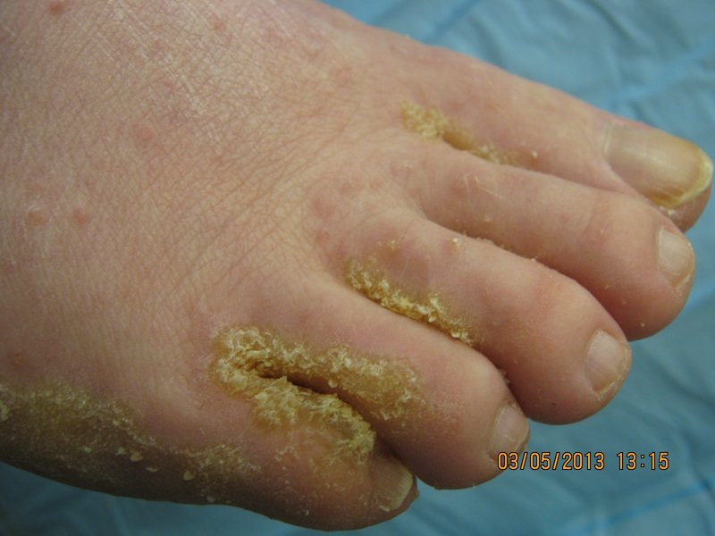
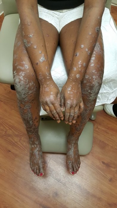
Clinical onset of keratosis follicularis usually arises in the third decade of life. The characteristic keratotic, yellowish brown warty papules and plaques often appear greasy and crusted.[4] Most prevalently, it is identified through examination of familial medical history. However, lack of an exclusive symptom may lead to a misdiagnosis of Darier’s disease as another dermatologic deformity. Clinical indicators of the disease may vary from very subtle, asymptomatic presentations to intense lesions that fluctuate with time, and are often chronic in nature.[5] The most commonly displayed symptoms of Darier’s disease are skin rashes, lesions affecting hands and nails, and lesions affecting mucous membranes. In approximately half of all cases of Darier’s disease, the oral mucosa is affected and most commonly discovered during routine dental appointment.[2] Resulting lesions of the disease form numerous stiff papules with normal, whitish, or reddish color that are most prevalent on the palatal and alveolar mucosa.
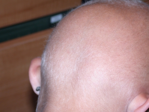
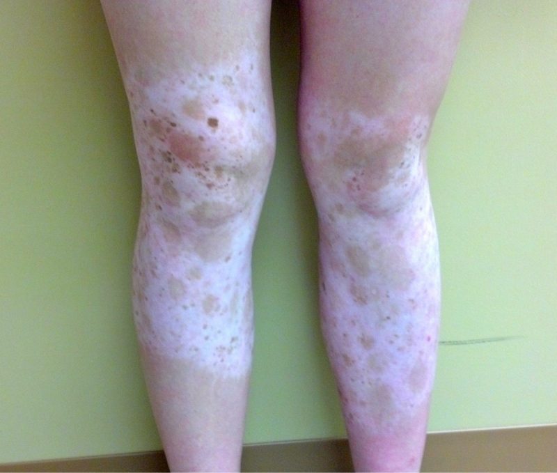
The unique segmental phenotypic consequences of Darier’s disease are believed to originate from a postzygotic mosaicism. Skin rashes and nail lesions often manifest symmetrically with hyperkeratotic, hyperpigmented papules and plaques in a seborrheic distribution.[6] However, as was evident in our patient’s case, the typical presentation may not perfectly correlate with the clinical picture. Similarly, another case of Darier’s disease identifies the elusiveness of this process. Due to minimal truncal involvement and a negative family history, this patient was misdiagnosed initially. The most critical symptom the individual displayed was extensive, hyperkeratotic verrucous plaques on the lower legs. Previous to this case, only four other medical reports noted this variant. A shave biopsy of the lower leg plaques confirmed the diagnosis.[7] Furthermore, Ferizi et al. reported a 53-year-old woman clinically displaying palmoplantar papules on the back of her hands and feet. The patient also suffered nail, nose, scalp, and ear deformity along with keratotic lower back and genital papules, and oral lesions forming white papules characterized by a central depression. Differential diagnosis of these symptoms led to six possible diagnoses including Darier’s disease. Again, only after biopsy was keratosis follicularis confirmed.[2]
TREATMENT:
Generally, only troublesome symptoms of Darier’s disease are treated. Mild symptomatic expression may be managed through the use of moisturizers, sunscreen, and proper awareness/use of clothing or conditioning that minimizes overheating, sweating, and mechanical trauma to affected regions. Furthermore, localized formations of the disease have successfully been treated to alleviate symptoms with the use of topical retinoids. As secondary bacterial or viral infection is a possible complication of keratosis follicularis, antibiotics and antivirals should be used if clinically indicated. In treating severe instances of Darier’s disease, oral retinoids have been used with success in the past. Due to possible side effects, a trial period with observation should be instituted at the appropriate dosage, as was recommended in our patient’s case. Acitretin and isotretinoin are currently at the forefront of this approach. Oral retinoids have shown to have the greatest impact on treating the disease and its symptoms, achieving alleviation in 90% of reported cases through reduction of hyperkeratosis and foul odor along with the smoothing of symptomatic papules.[5,8]
REFERENCES:
1) Kim, Carolyn. Keratosis follicularis (Darier-White disease), with an unusual palmoplantar keratoderma. Dermatology online journal 13.1 2007: 7. University of California, Davis.
2) Ferizi, Mybera. A Rare Clinical Presentation of Darier’s Disease. Case reports in
dermatological medicine 2013.6 2013: 419797. Hindawi Publishing.
3) Shi, Bing-Jun. The ATP2A2 gene in patients with Darier’s disease: one novel splicing mutation. International journal of dermatology 51.9 Sep 2012: 1074-7. Blackwell Publishing.
4) Chacon, Gina R., et al. “Darier’s disease: a commonly misdiagnosed cutaneous disorder.”Journal of Drugs in Dermatology 7.4 (2008): 387+. Academic OneFile.
5) Stanway, Amy. “Darier’s Disease.” DermNet NZ. NZDSI, 2003. Web.
6) Harboe, Theresa Larriba. Mosaicism in segmental Darier disease: an in-depth molecular analysis quantifying proportions of mutated alleles in various tissues. Dermatology (Basel) 222.4 2011: 292-6. S. Karger AG.
7) Katta, Rajani. Cornifying Darier’s disease. International journal of dermatology 39.11 Nov 2000: 844-5. Blackwell Publishing.
8) Kwok, Pui-Yan. “Keratosis Follicularis (Darier Disease).” Medscape. WebMD LLC, 21 Sept. 2012. Web.


