CORRECT DIAGNOSIS:
Leser-Trélat Sign
DISCUSSION:
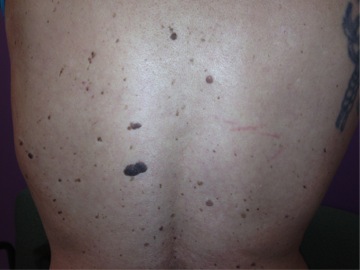
Edmund Leser and Ulysse Trélat, two European surgeons, first independently associated internal malignancies with these skin lesions in 1890. In addition, Eugen Holländer first associated “verrucae seborrheicae” with internal malignancy in 1900.1 Leser-Trélat Sign is a rare dermatological manifestation of a paraneoplastic syndrome characterized by a sudden abrupt onset of numerous seborrheic keratoses.2 The exact pathogenesis is unknown. Associated symptoms often include pruritus and velvety lichenified skin characteristic of acanthosis nigricans. Leser-Trélat Sign has previously been associated with the association of adenocarcinomas and lymphoproliferative disorders. Typically, the Leser-Trélat sign presents in the elderly,3 and has been associated with adenocarcinoma, most frequently of the colon, breast, or stomach, but also of the kidney, liver, and pancreas, among others.4 Although potentially paraneoplastic eruptive seborrheic keratoses in Leser-Trélat occurs in other organs and non-malignant ailments.5
Although the pathogenesis of Leser-Trélat Sign remains unclear, it may be related to an overproduction of growth hormone, epidermal growth factor (EGF), and transforming growth factor-alpha (α-TGF).6 The latter is not found in normal adult cells but in fetal cells and transformed cell lines. Both EGF and α-TGF bind to the EGF receptor found in keratinocytes, especially in actively proliferating cells of the basal layer of normal epidermis.7 Normally EGF receptors are present mainly on basal keratinocytes and decrease as keratinocytes differentiate in the upper epidermal layers. However, acanthosis nigricans, seborrheic keratoses, and acrochordons contain a dense population of receptors in all layers of the epidermis except for the stratum corneum.7 The similarity in hyperactivity of these growth factors during cutaneous malignancies and cutaneous paraneoplastic syndromes could provide the link between the expression of Leser-Trélat Sign due to squamous cell carcinoma. In countless cases, the removal of the malignancy greatly reduced the appearance of the Leser-Trélat Sign.3, 4, 7, 8
We report a case with a characteristic presentation of Leser-Trélat Sign associated with mediastinal squamous cell carcinoma. Although our case presentation is atypical, there are several cases of Leser-Trélat Sign in the literature with similar features. Heaphy et al. presented a case with a similar presentation, however in their case, the Leser-Trélat Sign was caused by adenocarcinoma of the lung, which is commonly associated with this cutaneous manifestation.4 In addition, Mukherjee et al. also described a case of squamous cell carcinoma of the lung with a paraneoplastic presentation. However, in that case, the paraneoplastic type was acanthosis nigricans.9 The variation in ailments that present with eruptive seborrheic keratosis makes the criteria for Leser-Trélat Sign a controversial topic in the medical community. Several studies were conducted which analyze the ailments of patients who present with eruptive seborrheic keratosis and compare them with a control group. These case-control studies revealed that there is no significant association of internal malignancies with eruptive seborrheic keratosis versus other ailments.10, 11 Although debated and disputed in multiple studies there continues to be a large database of literature justifying the sign of Leser-Trélat as a paraneoplastic syndrome. Regardless of the cause, physicians of all specialties should be aware of the presentation of the Sign of Leser-Trélat and its implications of malignancies, internal or cutaneous. This manifestation is a useful clue for undiagnosed malignancies and can facilitate treatment of the malignancy, therefore improving the prognosis.
TREATMENT:
Management and treatment per oncology.
REFERENCES:
Moore, R., & Devere, T. (2008). Epidermal manifestations of internal malignancy. Dermatologic Clinics, 26(1), 17-29. https://doi.org/10.1016/j.det.2007.09.002
Holdiness, M. R. (1986). The sign of Leser-Trélat. International Journal of Dermatology, 25, 564–572. https://doi.org/10.1111/j.1365-4362.1986.tb04697.x
Venegas, A., Felipe, V., Abudinén, A., Reydet, V., Brunie, V., & Arcuch, D. (2012). Signo de Leser-Trélat asociado a adenocarcinoma gástrico: Caso clínico. Revista médica de Chile, 140(12), 1585-1588. https://doi.org/10.4067/S0034-98872012001200010
Heaphy Jr, M. R., Millns, J. L., & Schroeter, A. L. (2000). The sign of Leser-Trélat in a case of adenocarcinoma of the lung. Journal of the American Academy of Dermatology, 43(2), 386-390. https://doi.org/10.1067/mjd.2000.104967
DeWitt, C. A., Buescher, L. S., & Stone, S. P. (2012). Cutaneous manifestations of internal malignant disease: Cutaneous paraneoplastic syndromes. In L. A. Goldsmith, S. I. Katz, B. A. Gilchrest, A. S. Paller, D. J. Leffell, & K. Wolff (Eds.), Fitzpatrick’s Dermatology in General Medicine (8th ed.). Retrieved from http://accessmedicine.mhmedical.com.ezproxylocal.library.nova.edu/content.aspx?bookid=392&Sectionid=41138876
Siedek, V., Schuh, T., & Wollenberg, A. (2008). Case report: Leser–Trelat sign in metastasized malignant melanoma. European Archives of Oto-Rhino-Laryngology and Head & Neck, 266(2), 297-299. https://doi.org/10.1007/s00405-008-0636-6
Ellis, D. L., Kafka, S. P., Chow, J. C., Nanney, L. B., & Inman, W. H. (1987). Melanoma, growth factors, acanthosis nigricans, the sign of Leser-Trélat, and multiple acrochordons: A possible role for alpha-transforming growth factor in cutaneous paraneoplastic syndromes. The New England Journal of Medicine, 317(25), 1582-1587. https://doi.org/10.1056/NEJM198712171172503
Ceylan, C., Alper, S., & Kılınç, I. (2002). Leser-Trelat sign. International Journal of Dermatology, 41, 687–688. https://doi.org/10.1046/j.1365-4362.2002.01600.x
Mukherjee, S., Pandit, S., Deb, J., Dattachaudhuri, A., & Bhuniya, S. (2011). A case of squamous cell carcinoma of lung presenting with paraneoplastic type of acanthosis nigricans. Lung India, 28(1), 62-64. https://doi.org/10.4103/0970-2113.76305
Fink, A., Filz, D., Krajnik, G., Jurecka, W., Ludwig, H., & Steiner, A. (2009). Seborrhoeic keratoses in patients with internal malignancies: A case–control study with prospective accrual of patients. Journal of the European Academy of Dermatology and Venereology, 23, 1316–1319. https://doi.org/10.1111/j.1468-3083.2009.03163.x
Lindelöf, B., Sigurgeirsson, B., & Melander, S. (1992). Seborrheic keratoses and cancer. Journal of the American Academy of Dermatology, 26, 947–950. https://doi.org/10.1016/0190-9622(92)70139-7


