CORRECT DIAGNOSIS:
Follicular Mucinosis
DISCUSSION:
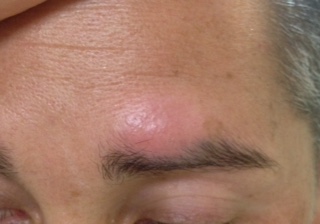
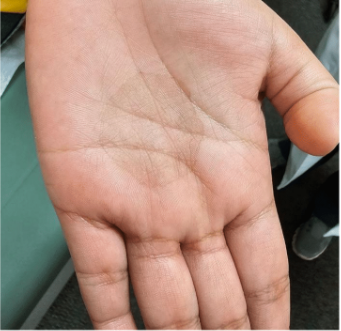
Alopecia mucinosis is a distinct entity described clinically as indurated or edematous follicular papule or plaques leading to hair loss. Lesion morphology is variable and can have a texture varying from smooth to scaly and lesions may be flesh-colored, white, or red. Lesions are most commonly found in the head and neck and are solitary, but lesions can be multiple especially if found on the body. Most lesions are asymptomatic, but the itching and loss of sensation have been reported.
The term primary follicular mucinosis is used to describe isolated skin lesions and carries a good prognosis, especially if lesions are solitary and the patient is under 40 years old.1 The disease tends to run a chronic, but benign course. Secondary follicular mucinosis is applied when the lesions are associated with malignancy such as mycosis fungoides or other inflammatory diseases such as systemic lupus erythematosus or atopic dermatitis. Recent reports have also documented follicular mucinosis associated with the medications adalimumab and imatinib mesylate.2,3 However, when lesions present in older patients or when there are multiple lesions than the patient’s condition most likely represents a cutaneous T-cell lymphoma (CTCL), or will progress to CTCL within 5 years.4 Clonal T-cell rearrangements can be found in be within both categories of follicular mucinosis, and its presence does not always represent a poor prognosis or future transformation to CTCL.1 No firm criteria can be used either clinically or histopathologically to differentiate primary follicular mucinosis from CTCL-associated follicular mucinosis so long-term follow up is always necessary.
The classic histopathologic descriptions of follicular mucinosis include excessive mucin deposition in the outer root sheath and sebaceous glands. A mixed inflammatory infiltrates may be found in the dermis of lesions. Atypical perifollicular infiltrate can be found when lesions are associated with CTCL, but epidermotropism is generally not found. Lymphoma may also be more likely (now, or in the future) if Pautrier’s microabscesses and significant lymphoid hyperplasia are found. T-cell receptor gene rearrangement may be found in both benign and malignant forms of follicular mucinosis.5,6,7
TREATMENT:
Follow up is essential for chronic disease and repeat biopsies are indicated to rule out the malignant transformation, but there are no firm guidelines on frequency of visits or specific examinations. In general, patients should be instructed to follow up immediately for any new or spreading lesions. Treatment is difficult and often unsatisfactory. Topical and intralesional steroids may improve or resolve individual lesions. Isolated case reports have shown success using isotretinoin, dapsone, psoralen plus UVA (PUVA), IFN alpha 2b, minocycline, and radiation therapy.8,9 Hydroxychloroquine appears promising with a case series showing cure in 6 patients, but more studies need to be before this is recommended as a first-line treatment.10 Our patient agreed to one treatment of 5 mg/cc intralesional Kenalog injection, but he has not returned for follow up.
REFERENCES:
1. Brown HA, Gibson LE, Pujol RM, et al.: Primary follicular mucinosis: long term follow-up of patients younger than 40 years with and without clonal T-cell receptor gene rearrangement. J Am Acad Dermatol. 2002;47:856-862.
2. Dalle S, Balme B, Berger F, Hayette S, Thomas L. Mycosis fungoides-associated follicular mucinosis under adalimumab. Br J Dermatol. Jul 2005;153(1):207-8.
3. Scheinfeld N. Imatinib mesylate and dermatology part 2: a review of the cutaneous side effects of imatinib mesylate. J Drugs Dermatol. Mar 2006;5(3):228-31.
4. Bi MY, Curry JL, Christiano AM, Hordinsky MK, Norris DA, Price VH, et al. The spectrum of hair loss in patients with mycosis fungoides and Sézary syndrome. J Am Acad Dermatol. Jan 2011;64(1):53-63.
5. Gerami P, et al.: The spectrum of histopathologic and immunohistochemical findings in folliculotropic mycosis fungoides. Am J Surg Pathol. 2007;31:1430.
6. Nickoloff BJ, Wood C. Benign idiopathic versus mycosis fungoides-associated follicular mucinosis. Pediatr Dermatol. Mar 1985;2(3):201-6.
7. Gibson LE, Muller SA, Leiferman KM, Peters MS. Follicular mucinosis: clinical and histopathologic study. J Am Acad Dermatol. Mar 1989;20(3):441-6.
8. Fernandez-Guarino M, Harto Castano A, Carrillo R, Jaen P. Primary follicular mucinosis: excellent response to treatment with photodynamic therapy. J Eur Acad Dermatol Venereol. Mar 2008;22(3):393-4.
9. Meissner K, Weyer U, Kowalzick L, Altenhoff J. Successful treatment of primary progressive follicular mucinosis with interferons. J Am Acad Dermatol. May 1991;24(5 Pt 2):848-50.
10. Schneider SW, et al.: Treatment of so-called idiopathic follicular mucinosis with hydroxychloroquine. Br J Dermatol. 2010;163:420.


