Presenter: Cherise Khani, DO
Dermatology Program: St. Barnabas Hospital
Program Director: Cindy Hoffman, DO
Submitted on: April 14, 2015
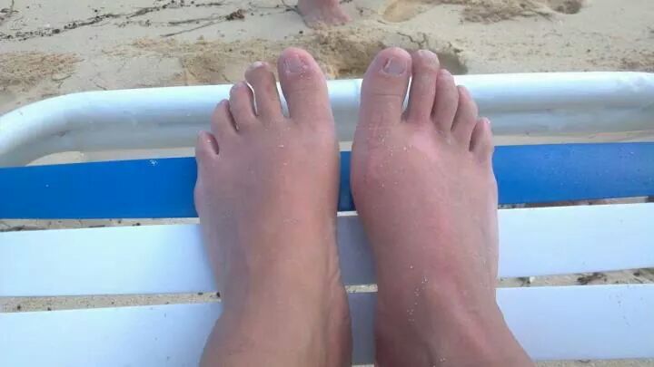
CHIEF COMPLAINT: Swelling of bilateral lower extremities x 2 years and “raised circles on right shin and toes” x 9 months
CLINICAL HISTORY: A 44 year-old female presented with swelling of her bilateral lower extremities x 2 years, and “raised circles on right shin and toes” x 9 months. Lesions of the right lower extremity were associated only with mild intermittent pruritus. The patient denied pain, numbness, or paresthesias of affected area. She was diagnosed with Graves’ disease in 2009 and successfully treated with radioactive iodine. Resultant hypothyroidism has been well controlled with levothyroxine, without recurrence of symptoms. The patient had a recent surgical excision of masses on right and left great toes, as well as degenerated sesamoid bone of the right foot. The surgical pathology demonstrated benign fibroconnective tissue, showing a myxoid-mucoid background.
Other information: Past medical history was significant for vulvar vestibulitis w partial vulvectomy, sinusitis w septoplasty, R foot bunion, GERD w nissen fundoplication, cervical dysplasia, and polyposis w LEEP, PCOS, chronic migraines, appendectomy, cholecystectomy, gastric bypass, and rotator cuff repair. Current medications consisted of Synthroid 150mg, oral contraceptive pill, Allegra, multivitamins, Glucosamine chondroitin, and Botox for migraines. Medication allergies included Versed, Fentanyl, Percocet, Morphine, Rocephin, Levaquin, and Vancomycin. Review of systems was otherwise negative
PHYSICAL EXAM:
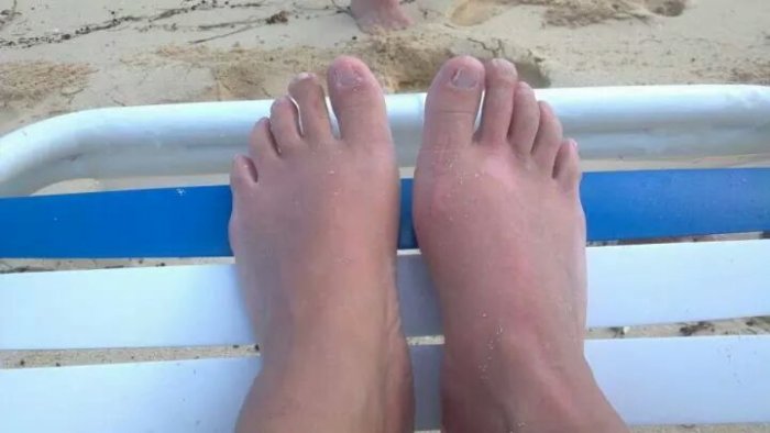
A skin type III female was found to have erythematous, indurated nodules on R shin and calf, with 3+ pitting edema of lower legs, ankles, and dorsal feet. The patient also had significant enlargement of bilateral great toes with overlying healed surgical incisions.
LABORATORY TESTS:
TSI 618 (H)
TSH 1.510 (WNL)
TPO 19 (WNL)
CBC, ESR, RF, CCP IgG, CRP WNL
HLA-B27 Ag and ANA NEG
DERMATOHISTOPATHOLOGY:
3.0 mm punch biopsy of right posterior distal leg: abundant mucin present within the reticular dermis
DIFFERENTIAL DIAGNOSIS:
1. Erythema nodosum
2. Lichen simplex chronicus
3. Hypertrophic lichen planus
4. Bullous pemphigoid (urticarial phase)
5. Lichen myxedematosus

