CORRECT DIAGNOSIS:
Toxic Epidermal Necrolysis
DISCUSSION:
Toxic epidermal necrolysis (TEN), aka Lyell’s Syndrome, is a rare potentially life-threatening drug reaction characterized by widespread mucocutaneous erythema, necrosis, and bullous detachment of the epidermis resulting in exfoliation(1).
TEN was first described by Debre et al in 1939 as l’erythrodermie bulleuses avec epidermolyse(2). In 1956 Alan Lyell presented four cases with an eruption resembling “scalding of the skin” and it was he who later coined the term “Toxic Epidermal Necrolysis”(1).
Worldwide, the average annual incidence of TEN is 0.4-1.2 cases per million population. In HIV positive populations the incidence of TEN increases 1000-fold3. Some risk factors for the development of TEN include HLA-B*1502 haplotype in Asians and East Indians exposed to Carbamazapine, HLA-B*5801 Han Chinese exposed to allopurinol and HLA-A*3101 Europeans exposed to Carbamazapine. In certain circumstances genotyping Asian individuals for the HLA-B*1502 allele is recommended prior to initiating therapy(1,4).
Use of medications is reported in 95% of patients diagnosed with TEN. Over 100 drugs have been implicated as a causative agent. Most frequently implicated medications include antibiotics (sulfonamides), anticonvulsants, NSAIDs, Allopurinol, and HAART therapies. The risk of developing TEN generally will occur within 7-21 days after taking such medications. Other more rare causes of TEN include infections and immunizations(1).
The pathogenesis of TEN involves an immune-related cytotoxic T-lymphocyte (CTL) reaction causing keratinocyte apoptosis and necrosis. It was discovered that these CTLs are activated via drug presentation by an antigen-presenting cell. Upon activation of CTLs, cytotoxic proteins, including granulysin, perforin/granzyme B, Fas ligand, and cytokines (INF-γ, TNF-α, IL-6 and IL-18) are produced which induce keratinocyte apoptosis. Notably, soluble fas ligand levels in the blood will directly correspond to body surface area involvement(1).
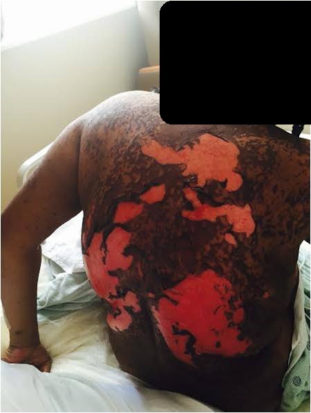
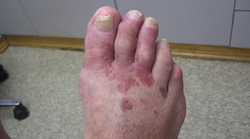
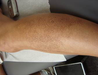
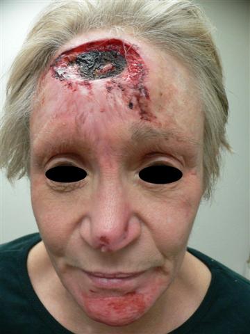
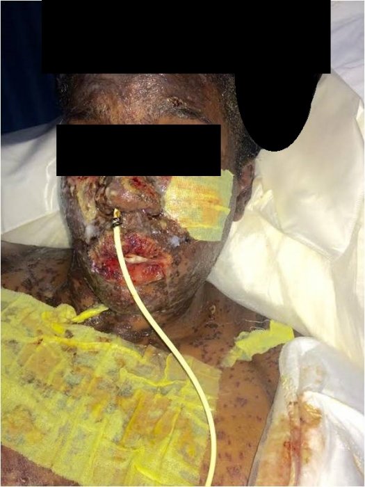
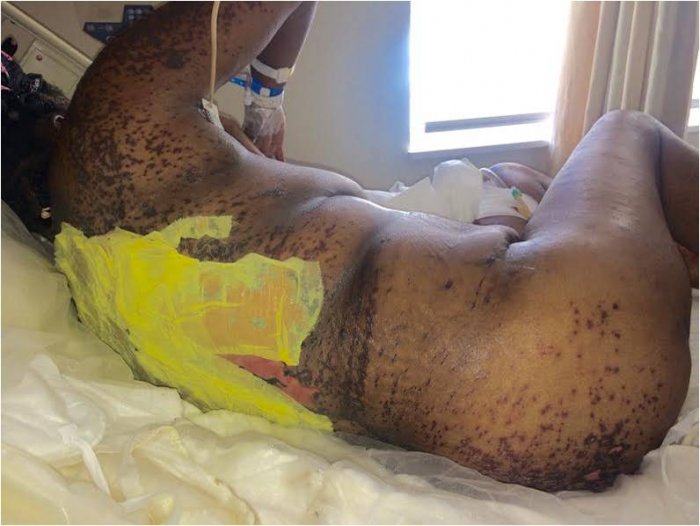
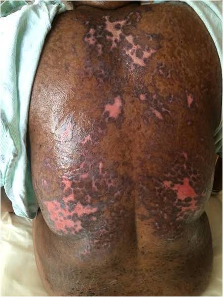
Upon initial presentation, patients may note fever, dysphagia, conjunctivitis or in our case, angioedema, preceding cutaneous rash by up to 3 days. Skin lesions usually start on the trunk and spread to face and extend caudally. Dusky red generalized macules are seen which eventually turn dusky followed by bullae and epidermal detachment. Histology shows a subepidermal blister with confluent necrosis of the entire epidermis with a sparse lymphocytic infiltrate. TEN is classified and differentiated from Stevens-Johnson Syndrome (SJS) based on body surface area (BSA) involvement with epidermal detachment(5).
• SJS < 10%
• SJS/ TEN overlap 10-30% BSA
• TEN > 30% BSA ( our patient )
• And then TEN without spots > 10% BSA
Mucous membrane involvement occurs and may result in gastrointestinal hemorrhage, respiratory failure, ocular abnormalities, and genitourinary complications. The mortality rate for TEN is 25-35%. Up to 35% of survivors will have long term ocular sequelae(1). Prognosis is guided by SCORTEN Criteria where 1 point is assigned for each prognostic factor below 1. Our patient had a SCORTEN of 1 throughout the hospital stay.
SCORTEN Prognostic Factors:(1)
• Age >40 years
• Presence of a malignancy (cancer)
• Heart rate >120 bpm
• Initial percentage of epidermal detachment >10%
• Serum urea level >10 mmol/L (28 mg/dL)
• Serum glucose level >14 mmol/L (252 mg/dL)
• Serum bicarbonate level <20
• SCORTEN 0-1 >3.2%
• SCORTEN 2 >12.1%
• SCORTEN 3 >35.3%
• SCORTEN 4 >58.3%
• SCORTEN 5 or more >90%
Treatment includes early diagnosis with immediate withdrawal of offending agent as well as supportive care.
TREATMENT:
Most importantly, the offending agent Lamictal was discontinued. Due to the late transfer of the patient to our facility, she was given a total of 1 day IVIG 1mg/kg/d. Xeroform dressings were applied to all areas of denuded skin. Hydrocortisone 2.5% ointment was applied to all non-denuded areas. Air mattress with pressure ulcer prophylaxis and nonstick sheets were utilized for bedding. Intravenous fluids ½ NS + 20 mEq KCL was started to keep urine output between 40-60 cc/hr. Of note, keeping TEN patients pre-renal will provide better patient outcomes and prevent fluid overload. Pain management was controlled with Morphine 4mg Q4 hours. A nasogastric tube was placed with progressively increased feeding volumes. Foley was placed as well. Tobradex ointment was applied to eyes Q6 hours with artificial tears placed intraocularly Q2 hours. Bactroban ointment was applied intranasally and periorally Q2hours. Gelfoam was eventually applied to lips for control of extensive mucosal bleeding. Lidocaine 5cc swish and spit was given Q2 hours and ½ strength peroxide to oral cavity BID. Whirlpool treatment was initiated 9 days after admission, once skin was re-epithelialized. The patient complained of genital pain on day 15 of admission and lidocaine gel and Acyclovir 500 mg Q8 hr x 7 days were prescribed at that time. Levaquin was given for the treatment of Enterococcus+ UTI. DVT prophylaxis on admission was initiated with Heparin 5000 unit subcutaneously BID which was eventually switched to Lovenox 80 mg BID after the unfortunate development of DVT on day 14 of admission. Protonix was given for GI prophylaxis with Zofran given as needed for nausea. The patient was discharged home on day 19 of admission with successful recovery.
REFERENCES:
1. Bolognia JL, Jorizzo JL, Rapini RP. In: Dermatology. 3rd ed. St. Louis (MO): Mosby Elsevier; 2012:324-333.
2. Debre R, Lemy M, Lamotte M. L’erythrodermie bulleuses avec epidermolyse. Bull Soc Pediatr (Paris). 1939;37:231-238.
3. Rzany B, Mockenhaupt M, Stocker U, et al. Incidence of Stevens-Johnson syndrome and toxic epidermal necrolysis in patients with the acquired immunodeficiency syndrome in Germany. Arch Dermatol. 1993; 129:1059.
4. Tangamornsuskan W, Chaiyakunapruk N, Somkrua R et al. Relationship between the HLA-B*1502 allele and carbamazepine-induced Stevens-Johnson syndrome and toxic epidermal necrolysis: a systematic review and meta-analysis. JAMA Dermatol. 2013 Sep;149(9):1025-32.
5. Bastuji-Garin S, Rzany B, Stern RS, Shear NH, Naldi L, Roujeau JC. Clinical classification of cases of toxic epidermal necrolysis, Stevens-Johnson syndrome, and erythema multiforme. Arch Dermatol. 1993 Jan. 129(1):92-6.

