CORRECT DIAGNOSIS:
Mycobacterium marinum
DISCUSSION:
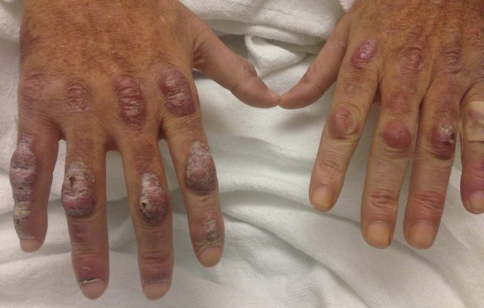
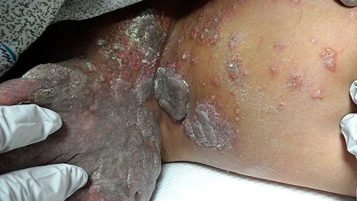
Mycobacterium marinum is a nontuberculoid acid-fast bacilli, which is a pathogen found in fish that has the rare potential to cause infection in humans.1-3 Infection requires exposure to contaminated water or direct contact with fish in conjunction with a skin break allowing for inoculation.1,2 The strong correlation of this infection with a marine environment has led to the following synonyms: aquarium granuloma, swimming pool granuloma and fish tank granuloma.3 The estimated incidence is 0.04-0.27 per 100,000.3 The most common location for infection is on the dorsal aspect of the hands as this is the site most at risk when handling fish and cleaning aquariums.1-4 Due to the indolent course and misdiagnosis as more common forms of cellulitis, the median time to diagnosis is three and a half months.1 Delayed diagnosis may increase the risk of invasive infection, leading to osteomyelitis and tenosynovitis.1,3 Diagnosis requires a high index of suspicion and a thorough history regarding travel history, hobbies, occupation, aquatic exposure and injury before onset. Patients present with an indolent course of erythematous, indurated papules and nodules with or without ulceration and exudate in an acral distribution. The lesions may follow a sporotrichoid pattern.1-4 Physical examination should include palpation of the lymph nodes draining the affected area. When Mycobacterium marinum is in the clinical differential diagnosis, two biopsies should be taken – one for histopathologic examination and one for tissue culture.1-4
Histopathologic examination will show a dermal suppurative and granulomatous process without caseation, as well as epidermal acanthosis and parakeratosis with or without ulceration. Although frequently negative, Ziehl-Neelsen and Fite staining should be applied to the tissue in an attempt to identify acid-bacilli.1-3,5 When sending the specimen in for tissue culture, it is important to work closely with your laboratory. Mycobacterium marinum will grow photochromic colonies on Lowenstein-Jensen medium with the optimum temperature between 86 and 91.4°F. Colonies become visible between 8-14 days and are white in the dark but become yellow-orange when exposed to light.4 Recent studies have shown that amplification of the hsp65 gene by PCR is an emerging tool used for rapid diagnosis of Mycobacterium marinum that is both sensitive and specific, but needs to be read with caution as false-positives have been reported.3 After tissue culture has been obtained, empiric antibiotic treatment with polymicrobial coverage is recommended. It is important to assess the patients Tetanus status as inoculation is often due to injuries occurring in a contaminated environment.2
Due to the rarity of this infection, there are no large randomized controlled studies assessing the efficacy of various treatment regimens. In simple cutaneous infections, monotherapy may be initiated with clarithromycin, minocycline, doxycycline or trimethoprim-sulfamethoxazole.1,3 Invasive infections must be treated with a combined treatment regimen. Clarithromycin in combination with an anti-tuberculoid medication, such as ethambutol or rifampin, is recommended. If there is an intolerance or allergy to the above medications, the addition of levofloxacin may be considered.1,3 There has been one recent case report describing a timely therapeutic response to lymecycline 150mg once daily. This may be a therapeutic consideration in the future due to the simple dosing, low risk of adverse effects, and gastrointestinal tolerability.4 Debridement is generally not recommended unless the lesions are refractory to treatment with the above antibiotic regimen.3 As a general rule, patients should be continued on antimicrobial therapy for one month after the resolution of clinical lesions.2
TREATMENT:
Due to the localized cutaneous nature of the infection, our patient has continued on monotherapy with trimethoprim-sulfamethoxazole 400-80 mg by mouth twice daily. She was also placed on silver sulfadiazine 1% topical cream applied to infection 1-2 times daily with dressing changes. At initial follow up appointment, lesions showed decreased induration and improvement of erythema. Complete resolution was seen after four months and the patient was instructed to continue trimethoprim-sulfamethoxazole until one month after lesions have resolved.
This case illustrates the importance of a thorough history and clinical examination. Conditions, such as Mycobacterium marinum, are easily overlooked or initially misdiagnosed when missing key elements of history. Delayed diagnosis can increase the risk of invasive infections and expose the patient to unnecessary antibiotic regimens.
REFERENCES:
(1) Johnson, M.G., & Stout, J.E. (2015). Twenty-eight cases of Mycobacterium marinum infection: Retrospective case series and literature review. Infection. Retrieved April 14, 2015.
(2) Baddour, L.M. (2015). Soft tissue infections following water exposure. UpToDate. Retrieved June 4, 2015.
(3) Sette, C.S., Wachholz, P.A., Masuda, P.Y., et al. (2015). Mycobacterium marinum infection: A case report. Journal of Venomous Animals and Toxins Including Tropical Diseases, 21, 7.
(4) Neugebauer, M.G., Neugebauer, S.A., Junior, H.L., et al. (2015). Treatment of Mycobacterium marinum with lymecycline: New therapeutic alternative? An Bras Dermatol, 90(1), 117-119.
(5) Belz, D., Tantcheva-Poor, I., Rasokat, H., et al. (2014). Mycobacterium marinum infection initially diagnosed as metastatic Crohn’s disease. Journal of the European Academy of Dermatology and Venereology, Letter to the Editor.


