Presenter: Danielle Raffaella Lazzara, DO
Dermatology Program: Larkin Community Hospital Palm Springs
Program Director: Brad Glick, DO, MPH, FAOCD, FAAD
Submitted on: April 27, 2018
CHIEF COMPLAINT: 1 year history of multiple, brown lesions diffusely spread on body
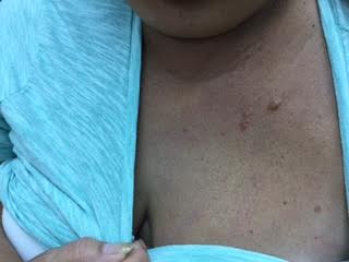
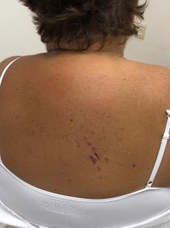
CLINICAL HISTORY: 66 year-old Hispanic female presented with a 1 year history of multiple, brown lesions located to the neck, chest, and upper back. The lesions were noted to be stable and asymptomatic with no aggravating factors. Patient denied fever, chills, arthralgia, weight loss, cough, shortness of breath, uveitis, back pain, abdominal/pelvic pain, hematuria, and dysuria. No previous treatment was performed. Patient’s medical history is significant for hypercholesterolemia managed medically with a statin and uterine fibroids for which she had a hysterectomy at age 33. She denies any pertinent family history.
PHYSICAL EXAM:
Physical examination was significant for multiple, firm, smooth-surfaced, pink-brown nodules, measuring 0.3 – 1.0 cm in diameter, located to the right mid-back, chest, and jawline. Lesions on the back were displayed in a linear distribution. The nodules were non-tender to palpation. Abdomen, extremities, groin, acral and mucosal sites were unaffected. No joint deformities or ocular abnormalities were noted.

LABORATORY TESTS:
No labs/ancillary studies were performed.
DERMATOHISTOPATHOLOGY:
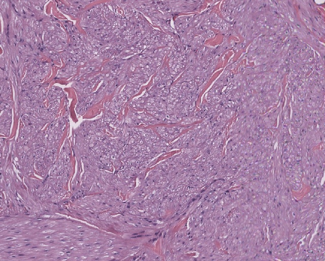
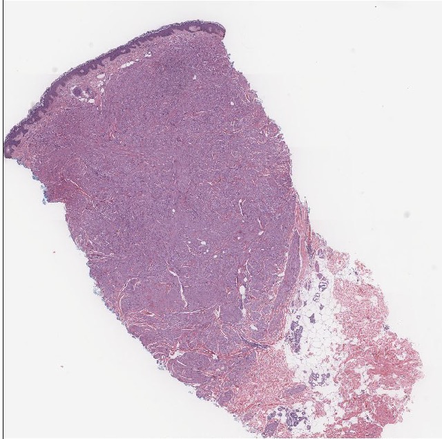
A 4.0 mm punch biopsy of lesion to the right back was performed. H&E displayed a non-encapsulated, spindle-cell neoplasm located to the reticular dermis composed of fascicles of elongated smooth muscle cells containing plump, blunt ended ‘cigar shaped’ nuclei. Nuclear atypia and mitotic activity not identified.
DIFFERENTIAL DIAGNOSIS:
1. Cutaneous Leiomyomas
2. Darier-Roussy Sarcoidosis
3. Neurofibromas
4. Subcutaneous Granuloma Annulare
5. Cutaneous Metastases


