Presenter: Helen Kaporis, D.O.
Dermatology Program: KCOM- Texas Division
Program Director: Dr Bill Way
Submitted on: March 4, 2011
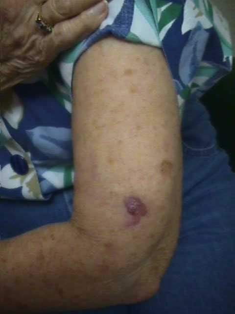
CHIEF COMPLAINT: Nodule of 6 months duration
CLINICAL HISTORY: Patient presented with an asymptomatic nodule of 6 months duration. No prior history of treatments to the lesion.
PHYSICAL EXAM:
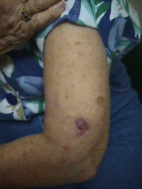
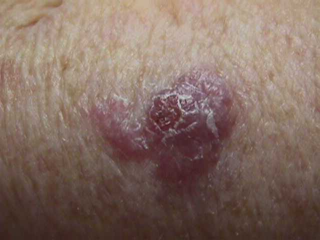
2 cm violaceous nodule of the left upper extremity
LABORATORY TESTS: N/A
DERMATOHISTOPATHOLOGY:
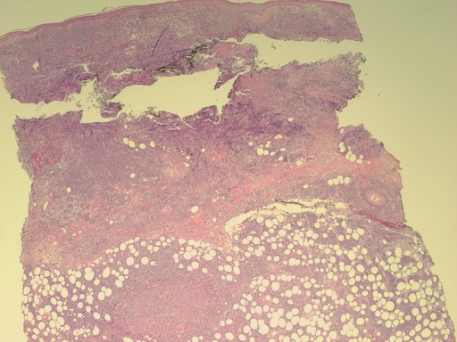
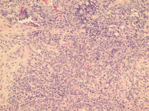
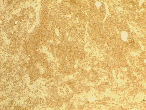
The diffuse nodular proliferation of markedly enlarged atypical lymphocytes involving the dermis and subcutaneous adipose tissue. The monomorphic population showed large nuclei and prominent nucleoli with scant cytoplasm. Abundant mitotic figures and pleomorphism were also noted. Epidermotropism was not observed. Immunohistochemical stains were positive for CD20, PAX5, and diffusely with BCL-2, as well as BCL-6, CD-79a, and slightly for CD10. Ki-67 immunostain highlighted a large percentage of the tumor cells.
DIFFERENTIAL DIAGNOSIS:
1. Basal Cell Carcinoma
2. Kaposi’s Sarcoma
3. Lymphoma
4. Leishmaniasis

