Presenter: Eric Sandrock, DO; Rick J H Lin, DO
Dermatology Program: South Texas Dermatology Residency Program, HCA Corpus Christi Medical Center Bay Area
Program Director: Rick Lin, DO
Submitted on: December 10, 2024
CHIEF COMPLAINT: Persistence of pigmented lesion on forehead
CLINICAL HISTORY: A 73-year-old female visited the dermatology clinic for a routine skin evaluation. She had no personal history of skin cancer but reported noticing changes in the appearance of some lesions. During the visit, five shave biopsies were performed. After receiving concerning pathology results, an excision was scheduled, but the patient did not return for 37 months. Upon her return at age 76, the patient was re-evaluated, and it was noted that the previously biopsied pigmented lesion on her right frontal hairline had enlarged.
PHYSICAL EXAM:
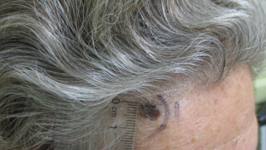
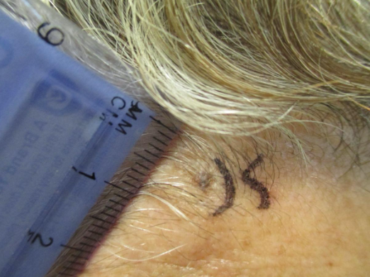
Upon the initial examination, the patient showed signs of widespread sun damage, along with several scattered lesions that varied in elevation and pigmentation. Most concerning was a 3mm melanocytic lesion with variable color and irregular border on the right frontal hairline.
Upon re-evaluation 37 months later, the melanocytic lesion on the right frontal hairline measured 5 x 10 mm. It exhibited a deeper pigmentation compared to the initial biopsy and displayed irregular borders.
LABORATORY TESTS: N/A
DERMATOHISTOPATHOLOGY:
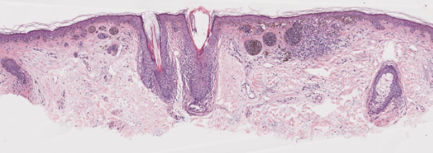
Initial biopsy stained with hematoxylin and eosin, showing plump melanocytes arranged in nests, surrounded by mixed inflammatory cells occupying the upper dermis. Solar elastosis is also noted at the base of the specimen.
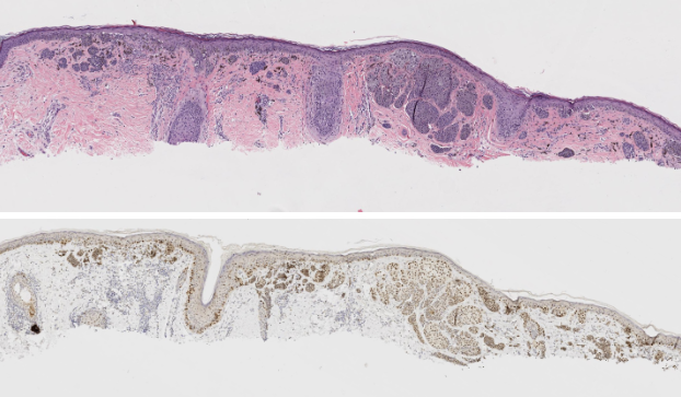
Re-biopsy stained with hematoxylin and eosin (top) as well as MART-1 staining (bottom) to confirm the melanocytic lesion. The microscopic evaluation showed evidence of radial growth phase, with fewer than 1 mitotic figure per mm squared. There was no ulceration, regression, vascular or lymphatic invasion, or perineural invasion. The melanocytes were predominantly epithelioid, with the lesion extending to the deep margin of the specimen and involving the peripheral margins and adnexal structures. Atypical melanocytes present within the upper dermis, at a Breslow depth of at least 0.6 mm.
DIFFERENTIAL DIAGNOSIS:
- Compound Melanocytic Nevus
- Melanoma in situ
- Malignant Melanoma
- Basal Cell Carcinoma
- Squamous Cell Carcinoma
- Seborrheic Keratosis
- Angiokeratoma
- Kaposi Sarcoma

