Presenter: Eugene Sanik DO, Jordan Fabrikant DO
Dermatology Program: Larkin Community Hospital / NSUCOM
Program Director: Stanley Skopit DO, MSE, FAOCD
Submitted on: September 23, 2013
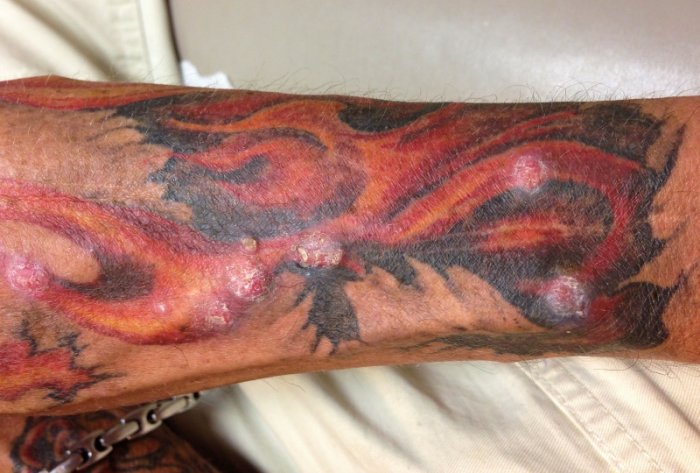
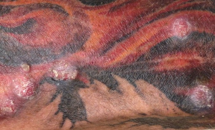
CHIEF COMPLAINT: Multiple asymptomatic nodular lesions confined to the red areas of a new multicolored tattoo located on the left forearm
CLINICAL HISTORY: A healthy 57-year-old Caucasian male presents with multiple asymptomatic nodular lesions confined to the red areas of a new multicolored tattoo located on the left forearm. The tattoo was made two weeks earlier by the same professional artist who had done several tattoos for our patient in the past. Past medical history was negative for skin cancers and immunosuppression. Social history was significant for the patient who has been a World Trade Center rescuer with the New York Fire Department.
PHYSICAL EXAM:
On examination, we noticed nine firm hyperkeratotic, verrucous, and dome-shaped papules and nodules arranged in a curvilinear pattern corresponding to the sites of red pigment along with a flame tattoo. Older professional multicolored tattoos were present on both upper extremities but did not have any lesions present within them. Signs of photoaging and chronic sun exposure were observed on sun-exposed areas of the body including the face and extensor hands and forearms.
LABORATORY TESTS: N/A
DERMATOHISTOPATHOLOGY:
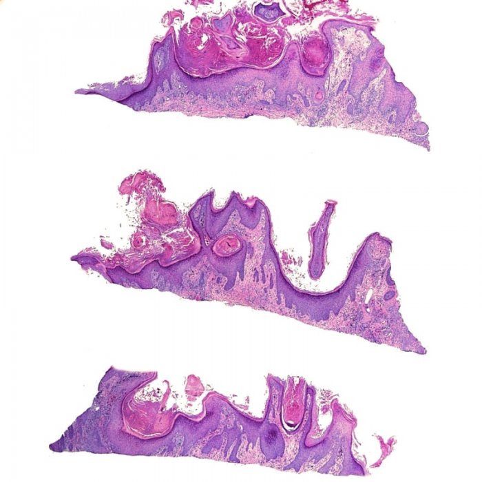
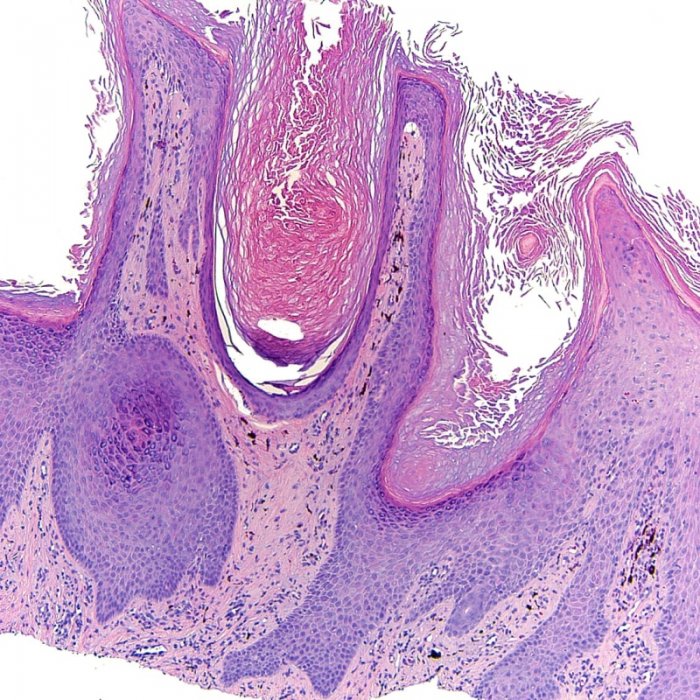
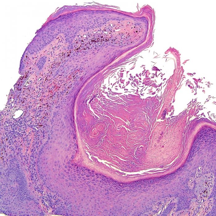
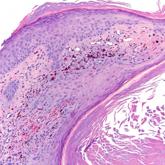
Skin biopsy was performed, and the histopathology showed a crateriform exo-endophytic growth with broad irregular epidermal hyperplasia and central hyperkeratosis. Higher magnification revealed squamous cells with variably glossy cytoplasm and exogenous tattoo pigment in the underlying dermis, without granulomatous reaction. A very small amount of tattoo pigment was seen within the central keratin plug undergoing transepidermal elimination.
Gram stain was negative for pathogenic bacteria. Acid-fast bacilli staining and tissue culture were negative for mycobacterium. Gomori methenamine silver (GMS) staining was negative for fungi.
DIFFERENTIAL DIAGNOSIS:
1. Foreign Body Granulomas
2. Eruptive Keratoacanthomas
3. Cutaneous Sarcoidosis
4. Atypical Mycobacterium
5. Inoculation of Viral Warts


