Presenter: Jacqueline Thomas, DO; Leeor Porges, DO; Khasha Touloei DO; Paloma Reiter, BA
Dermatology Program: Broward Health/Nova University
Program Director: Tracy Favreau DO
Submitted on: October 6, 2014
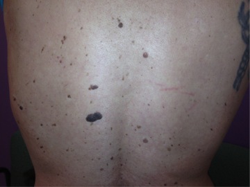
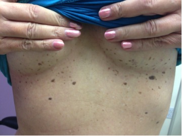
CHIEF COMPLAINT: several new lesions on back and inframammary region
CLINICAL HISTORY: A 59-year-old Caucasian female, with Fitzpatrick type III skin, with a history of hemochromatosis, Ehlers-Danlos syndrome, molar pregnancy, COPD, osteoarthritis, chronic pain syndrome, and chronic fatigue. The patient presented with several new lesions of concern on her back and inframammary region. No previous treatments.
PHYSICAL EXAM:
The lesions were numerous stuck on plaques that were waxy brown to dark brown.


Dermoscopically they presented with comedone like openings, milia like cysts, and bulbous projections. The lesions were first noted nine years prior and appeared rapidly. The patient denied a change in size and color, however, she denies pruritus. In addition, she also displayed generalized hair thinning and non-scarring alopecia of the scalp. On the remaining physical exam, the patient appeared generally thin, with aphasia, but no palpable lymphadenopathy was appreciated.
LABORATORY TESTS:
The patient initially sought medical assistance in September 2012 for aphasia. A CT of the neck and chest noted a hypodense nodule in the lower left lobe of the thyroid and a 3.8 cm left anterosuperior mediastinal mass and adenopathy. Biopsies of both lesions determined that the thyroid nodule was benign however the mediastinal mass was consistent with squamous cell carcinoma. The patient was found to have a persistent mediastinal squamous cell carcinoma with a non-small cell squamous cell carcinoma of the lung. The patient was classified as (TxN2M0 stage IIIa). The mass effect caused aphasia secondary to left vocal cord paralysis resulting from recurrent laryngeal nerve involvement. The mass was treated with stereotactic body radiation therapy (SBRT) and the patient underwent CyberKnife radiosurgery in five fractions. The patient was not offered chemoradiation therapy nor neoadjuvant chemotherapy. In May 2013 a PET scan identified that the mediastinal mass reduced to 2.5 cm in size
The follow-up PET scan in November 2013 noted a hypermetabolic lesion in the region of the aortic arch; with no change from prior imaging. In addition, the patient had an elevated CEA of 11.52 ng/ml. CT of the chest in February 2014 demonstrated a gradual increase of the left mid lung zone mass that measured 3.5 x 2.5 cm. A stable subcentimetric nodular density along the lateral aspect of the left major fissure was noted as well. No abnormal hilar or mediastinal adenopathy was noted but it was subsequently determined that the malignancy was unresectable.
A CT guided biopsy and Theros CancerType ID noted a primary keratinizing squamous cell carcinoma of the lung, with necrosis and calcifications. Further evaluation included whole body radiological imaging, and results showed no additional areas of focus or abnormality. The patient denied palliative chemotherapy and elected to be closely monitored. Lastly, she could not correlate the exact onset of her seborrheic keratoses with her malignancy, although she denied significant keratotic lesions prior to her diagnosis.
DERMATOHISTOPATHOLOGY:
Two lesions suspicious for atypical processes were biopsied, by shave removal, from the mid-back and left submammary regions. The biopsy revealed the diagnoses of seborrheic keratoses. Several other lesions on the back and submammary region suspicious for irritated benign growths were treated with cryotherapy.
DIFFERENTIAL DIAGNOSIS:
1. Benign seborrheic keratoses
2. Melanoma
3. Epidermal nevi
4. Pigmented BCC
5. Melanocytic nevi


