Presenter: Debbbie Palmer, DO and Dimitria Papadopoulos, DO, 2nd year residents
Dermatology Program: St. Barnabas Hospital Dermatology Department, Bronx, New York
Program Director: Cindy Hoffman, DO, FAOCD
Submitted on: May 29, 2002
CHIEF COMPLAINT: bilateral lower extremity edema and a growing red nodule
CLINICAL HISTORY: A 45-year-old Black male was presented from the nursing home with a one-week history of bilateral lower extremity edema and a few months of a nonpruritic, progressively enlarging growth on his left foot. This growth has bled with mild trauma. His past medical history is significant for HIV, endocarditis, cardiomegaly, congestive heart failure, intravenous drug abuse, end-stage renal disease, and pneumonia. The patient has not received any previous treatment for his current condition. His medication regimen includes methadone, temazepam, zolpidem, calcium carbonate, calcitriol, folic acid, a multivitamin, ferrous sulfate, and trimethoprim-sulfamethoxazole.
PHYSICAL EXAM:
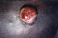
The patient was afebrile and was found to have bilateral lower extremity pitting edema that was tender and warm to touch, and a 1 centimeter, well-circumscribed, hemorrhagic, nodule on the left medial foot.

LABORATORY TESTS:
1. CBC- leukocyte count of 7.3 x 10³/UL (normal, 4.8 to 11 x 103/UL), polymorphonuclear leukocytes 35.9% (normal, 40 to 74%), lymphocytes 49.8% (normal, 19 to 48%), basophils 2.4% (normal, 0 to 1.5%), hemoglobin of 8.9 g/dL (normal, 14 to 18 g/dL), hematocrit of 27.5% (normal, 42 to 52%), platelets of 224 x 103/UL (normal, 130 to 450 x 103/UL)
2. Hepatic Function Panel- Within normal limits
3. T4 lymphocyte count- 107 cumulative cells (normal, 400 to 1770 cells/cumm)
4. T8 lymphocyte count- 2292 cumulative cells (normal, 240 to 1200 cells/cumm)
5. Absolute lymphocyte count- 2665 (normal, 960 to 4320 cells/cumm)
6. Blood cultures- no growth of any organisms
7. Shave biopsy of lesion on left medial foot
8. Pelvic and retroperitoneal ultrasounds- negative
9. Bilateral extremity doppler ultrasounds- negative
Bilateral x-rays of the feet- negative
DERMATOHISTOPATHOLOGY:
Microscopic Description: an atrophic epidermis with pseudo-epitheliomatous hyperplasia and lobular proliferations of small blood vessels containing cuboidal endothelial cells. The inflammatory cell infiltrate surrounding the vessels consisted of neutrophils. A Warthin-Starry silver stain was positive.
DIFFERENTIAL DIAGNOSIS:
1. Pyogenic granuloma
2. Kaposi’s sarcoma
3. Papular angiokeratoma
4. Bacillary angiomatosis


