CORRECT DIAGNOSIS:
Bacillary angiomatosis
DISCUSSION:
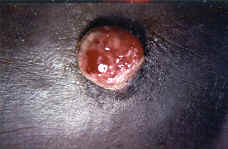
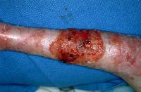
We describe a patient with an enlarging hemorrhagic nodule on the left medial foot. An initial diagnosis of cellulitis was made and the patient was treated with a course of amoxicillin. After thorough clinical and histological examinations, a diagnosis of bacillary angiomatosis was made. Bacillary angiomatosis is an angioproliferative disease that was first described in 1983 by Stoler et al.1,2 It is often described in severe immunodeficiency, as in advanced acquired immunodeficiency syndrome.2,3 Bacillary angiomatosis has derived its name from the vascular proliferation seen in histological specimens and from the presence of numerous bacillary organisms detected with Warthin-Starry silver stain or electron microscopy.2
Although Bartonella infections are not uncommon, bacillary angiomatosis remains a rare disorder with 1.2 cases per 1000 HIV infected patients.2 It has been observed that those most affected are patients with CD4 cell counts less than 100/ul.2
The first reported incident of infection with organisms of Rochalimaea genus, now known as Bartonella, was during World War I and was termed trench fever.2 The transmission occurred through the bite of the human body louse.5 In 1990, Relman et al. recognized a relationship between trench fever and Rochalimaea Quintana, now known as Bartonella Quintana, the cause of bacillary angiomatosis.2,6 There is an association between B Quintana infections and homelessness, due to transmission through the body louse vector.2 In 1992, another member of the genus Bartonella, B Henselae, was also recognized as another etiological agent of this illness.2 It is transmitted to humans through direct contact with cats or the cat flea, Xenopsilla cheopis.2 B Quintana is responsible for most cases of subcutaneous infection, deep soft tissue disease, and lytic bone lesions.5 Liver and lymph node involvement has been associated with B Henselae infection.5 Bartonella species are fastidious gram-negative bacteria.3
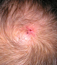
Bacillary angiomatosis typically presents as an inflammatory disease, most often involving the skin.2 The skin lesions are either single or multiple and can be localized in cutaneous and subcutaneous tissues.2 The usual lesion is an angiomatous papule or nodule, resembling a pyogenic granuloma or a subcutaneous nodule.7 The atypical lesions can present as Kaposi’s sarcoma or papular angiokeratoma.7 Other areas of involvement are the oral, anal, conjunctival, and gastrointestinal mucosal surfaces, brain, respiratory tract, liver, spleen, bone, bone marrow, and lymph nodes.2,5 Lesions evolve slowly over a period of several weeks and patients usually complain of fever and weight loss, chronic lymphadenopathy, and abdominal pain.2,4
The diagnosis of bacillary angiomatosis is established by clinical and histological examination of affected tissues.2 Culture of Bartonella species from serum remains insensitive and very few isolates are available worldwide.4 Polymerase chain reaction amplification is another method used in detecting Bartonella species in biopsy specimens.4
A high level of clinical suspicion remains the most important first step in diagnosing bacillary angiomatosis in immunocompromised patients. Recognition of bacillary angiomatosis is important because it can be cured with appropriate antibiotic therapy and can be life-threatening if left untreated.4,5
TREATMENT:
After a definitive diagnosis, our patient’s antibiotic regimen was changed. The patient was treated with erythromycin, 2 grams a day, and had subsequent clearing of the hemorrhagic nodule, after 8 weeks of therapy. Erythromycin, 2 grams a day, is the treatment of choice, while doxycycline, 100 mg twice a day, is used as an alternative therapy.2,4 Treatment should be continued for 2 to 3 months in an immunocompromised patient.2,4 It is not uncommon to have a relapse of bacillary angiomatosis upon withdrawal of antibiotic therapy.4 Precautionary measures in immunocompromised patients should include avoidance of contact with cats, fleas, and lice.4 Permethrin 1% dusting powder is the drug of choice for delousing and should be applied to clothing and bedding.4
REFERENCES:
Stoler, M. H., Bonfiglio, T. A., Steigbigel, R. T., & Pereira, M. (1983). An atypical subcutaneous infection associated with acquired immune deficiency syndrome. American Journal of Clinical Pathology, 80(6), 714–718. https://doi.org/10.1093/ajcp/80.6.714 [PMID: 6418072]
Plettenberg, A., Lorenzen, T., Burtsche, B. T., Rasokat, H., Kaliebe, T., et al. (2000). Bacillary angiomatosis in HIV-infected patients: An epidemiological and clinical study. Dermatology, 201(4), 326–331. https://doi.org/10.1159/000018086 [PMID: 11089506]
La Scola, B., & Raoult, D. (1999). Culture of Bartonella quintana and Bartonella henselae from human samples: A 5-year experience (1993 to 1998). Journal of Clinical Microbiology, 37(6), 1899–1905. https://doi.org/10.1128/JCM.37.6.1899 [PMID: 10364587]
Gasquet, S., Maurin, M., Brouqui, P., Lepidi, H., & Raoult, D. (1998). Bacillary angiomatosis in immunocompromised patients. AIDS, 12(14), 1793–1803. https://doi.org/10.1097/00002030-199814000-00014 [PMID: 9740171]
Santos, R., Cardoso, O., Rodrigues, P., Cardoso, J., Machado, J., et al. (2000). Bacillary angiomatosis by Bartonella quintana in an HIV-infected patient. Journal of the American Academy of Dermatology, 42(2), 299–301. https://doi.org/10.1016/S0190-9622(00)90011-1 [PMID: 10684443]
Relman, D. A., Loutit, J. S., Schmidt, T. M., Falkow, S., & Tompkins, L. S. (1990). The agent of bacillary angiomatosis: An approach to the identification of uncultured pathogens. New England Journal of Medicine, 323(22), 1573–1580. https://doi.org/10.1056/NEJM199011293232203 [PMID: 2239756]
Schwartz, R. A., Nychay, S. G., Janniger, C. K., & Lambert, W. C. (1997). Bacillary angiomatosis: Presentation of six patients, some with unusual features. British Journal of Dermatology, 136(1), 60–65. https://doi.org/10.1046/j.1365-2133.1997.12938.x [PMID: 9011330]


