CORRECT DIAGNOSIS:
Tinea nigra
DISCUSSION:
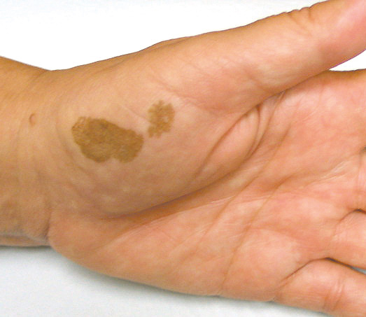
Tinea nigra is a rare superficial fungal infection of the palmar and plantar stratum corneum. Currently identified as Exophiala phaeoannellomyces, this dermatophyte had a previous taxonomic designation of Exophiala werneckii. Other dematiaceous fungi have been linked with the disease, including Stenella araguata, although Exophilia phaeoannellomyces is considered the most common cause. Tinea nigra infection clinically appears as mottled, brownish-black, velvety macules with fine-scale arising on the palms, volar aspect of fingers, or soles. Lesions are asymptomatic and over time increase in size and darken in appearance. Tinea nigra is most common in tropical regions such as South or Central America but has been reported in the United States particularly in Florida, Texas, and North Carolina. The fungus is ubiquitous to the soil, sewage, or decaying vegetation. Infection occurs most commonly by inoculation, although rare human to human transmission has been described. Incubation time can range from weeks to years.
Diagnosis can be easily established by the KOH examination of a lesional scraping. Brown to green hyphae and budding yeast cells are seen. Characteristically, hyphae are freely branching and septate, whereas yeast cells are single or paired and oval to spindle in shape. Mycological isolation initially reveals yeast-like colonies that appear brown to shiny-black in color. As cultures age, filamentous colonies with peripheral aerial mycelium develop, creating a grayish-green appearance. Biopsy typically yields subtle changes consisting of mild hyperkeratosis with a sparse dermal lymphocytic infiltrate. Exocytosis of neutrophils, typical of dermatophytosis, is not conspicuous features of this entity. Although pigmented spores and branched hyphae can be seen with high power scrutiny of the stratum corneum, these elements are more readily identified by GMS or PAS staining.
TREATMENT:
This patient began a three-week course of topical econazole and reported complete resolution without recurrence.
Topical treatment remains to be the primary management of Tinea nigra. Antifungals from the imidazole family (clotrimazole, miconazole, ketoconazole, sulconazole, econazole) are most effective. Treatment duration of at least 2 to 3 weeks is recommended. Alternative therapies include shaving of the superficial epidermis with a no. 15 blade alone or in combination with topical keratolytics and antifungal preparations (Whitfield’s ointment, topical thiabendazole, and retinoic acid). Griseofulvin and topical tolnaftate have been ineffective in treatment along with oral terbinafine therapy; however, topical terbinafine was reported successful in clearing lesions.
REFERENCES:
Elgart, M. L. (Ed.). (1996). Dermatologic clinics (Vol. 14). Philadelphia: Saunders.
Fader, R. C., & McGinnis, M. R. (1988). Infections caused by dematiaceous fungi: Chromoblastomycosis and phaeohyphomycosis. Infectious Diseases Clinics of North America, 2(4), 925–938. https://doi.org/10.1016/S0891-5520(18)30039-4
Rippon, J. W. (1988). Superficial infestations: Tinea nigra. In Medical mycology: The pathologic fungi and the pathogenic actinomyces (3rd ed.). Philadelphia: W.B. Saunders.
Babel, D. E., Pelachyk, J. M., & Hurley, J. P. (1986). Tinea nigra masquerading as acral lentiginous melanoma. Journal of Dermatologic Surgery and Oncology, 12(5), 502–504. https://doi.org/10.1111/j.1524-4725.1986.tb00257.x
Sayegh-Carreno, R., et al. (1989). Therapy of tinea nigra plantaris. International Journal of Dermatology, 28(1), 46–48. https://doi.org/10.1111/j.1365-4362.1989.tb02015.x


