Presenter: Michael Sabat, MS, DO, Megan Goff, DO, and Sam Smith, MPH, DO
Dermatology Program: Stephen Kessler, D.O.
Program Director: Stephen Kessler, D.O.
Submitted on: October 29, 2002
CHIEF COMPLAINT: Painful blisters/ ulcerations since birth
CLINICAL HISTORY: Patient presented with fragility of skin and slow healing, scarring erosions and ulcers, difficulty swallowing, decreased appetite and weight loss. Previous treatments include oral antibiotics, pureed diet, high-calorie diet, wound care including Vaseline gauze and Bactroban ointment, TAC 0.1% ointment, mineral oil, senicot, and iron. Of note, 12-year-old sister has a similar presentation. Neither the parents nor other siblings are affected.
PHYSICAL EXAM:
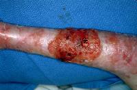
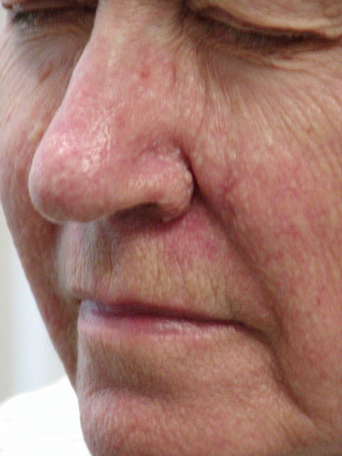
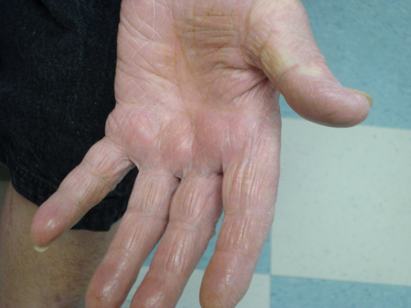
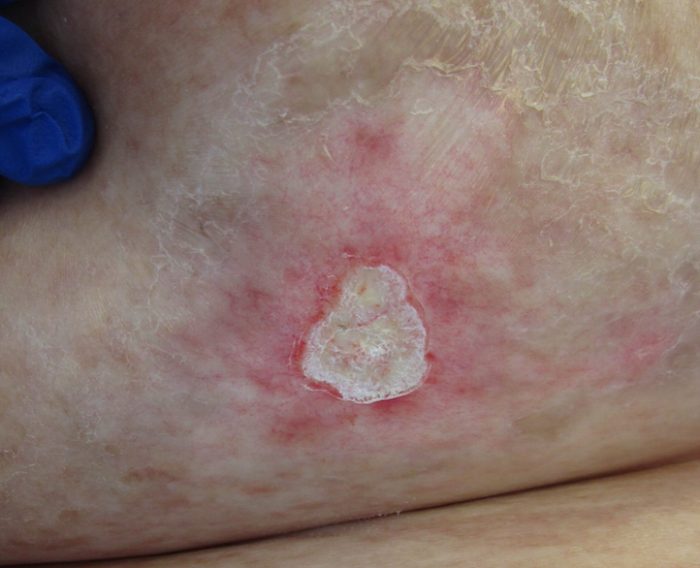
Thin cachectic appearing 11-year-old female appears younger than her stated age. Erosions, ulcerations, and scarring of the neck, posterior shoulders, axilla, lower abdomen, and scalp. Pseudosyndactyly of the 1st and 2nd digits of bilateral feet with anonychia. Scarring of the palms, soles, and scarring alopecia of the temporal scalp. Oral mucosa with small linear erythematous erosions on the soft palate.

LABORATORY TESTS:
Iron Deficiency Anemia on CBC and Iron Studies.
Positive Electron Microscopy showing absent anchoring fibrils.
Blood Leukocyte DNA analysis positive for COL7A1 gene mutations.
DERMATOHISTOPATHOLOGY:
Microscopic description: Monoclonal antibody against Collagen 7 demonstrates the absence of Collagen type 7 in the lesional, near perilesional, and distant perilesional skin. Cleavage plane of blistering is below the lamina densa by staining for BP230 and Collagen type IV which both localize to blister roof.
DIFFERENTIAL DIAGNOSIS:
1. Epidermolysis Bullosa Simplex
2. Junctional Epidermolysis Bullosa
3. Recessive Dystrophic Epidermolysis Bullosa
4. Dominant Dystrophic Epidermolysis Bullosa


