Presenter: Dan J. Ladd, DO, Rick Lin DO MPH
Dermatology Program: Texas Division of KCOM Dermatology Residency Program
Program Director: Bill V. Way, DO
Submitted on: July 30, 2003
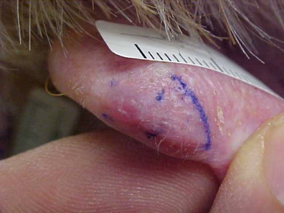
CHIEF COMPLAINT: Lesion on the left ear that has been slowly growing x 1 year
CLINICAL HISTORY: A 74-year-old Caucasian male presents with a lesion on the left ear that has been slowly growing for 1 year. The lesion is asymptomatic, does not bleed or ulcerate, and has had no previous treatment.
PHYSICAL EXAM:
Left ear helix at 12 o’clock reveals a solitary 7mm firm pearly dome-shaped papule with telangiectasias and slight central umbilication. No ulceration or scale.
LABORATORY TESTS: N/A
DERMATOHISTOPATHOLOGY:
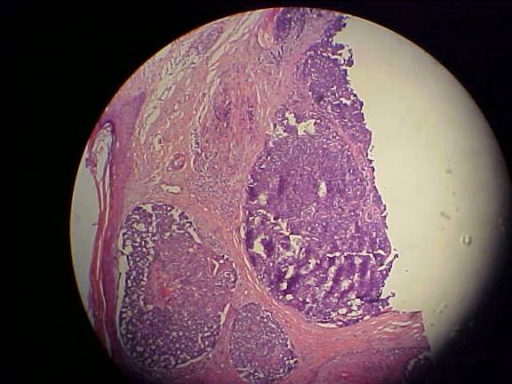
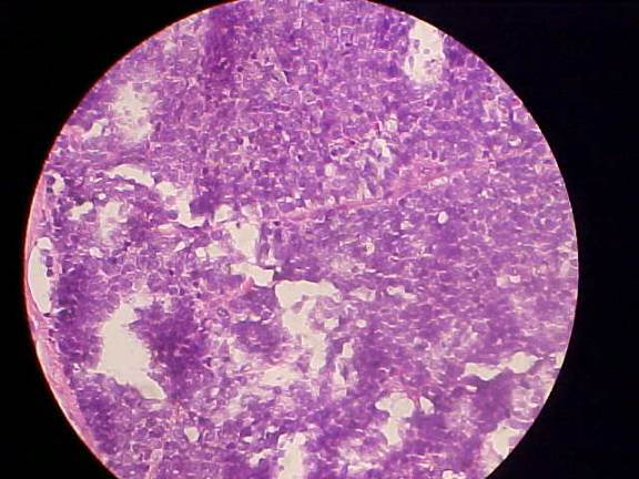
Poorly differentiated basophilic neoplasm characterized by sheets of atypical basophilic cells, many of which are necrotic and in mitoses. Special stains show that the cells are positive for cytokeratin 20 and neuron-specific enolase. See low power and high power digital photomicrographs below.
DIFFERENTIAL DIAGNOSIS:
1. Infiltrative basal cell carcinoma
2. Merkel cell carcinoma
3. Malignant eccrine spiradenoma
4. Desmoplastic trichoepithelioma
5. Adenoid cystic carcinoma


