CORRECT DIAGNOSIS:
Erythema annulare centrifugum (EAC)
DISCUSSION:
EAC is believed to be a hypersensitivity reaction to an underlying inflammatory or neoplastic disease, either benign or malignant. An eruption may wax and wane paralleling the primary process. EAC and EGR are the two reactive erythemas associated with malignancy. EGR, however, is more closely associated with neoplasia, with only a minority of cases reported in patients with EAC. When a malignancy is discovered in association with EAC, however, no particular type of cancer appears to predominate. In many cases, the EAC resolved following treatment of the tumor. EAC has also been reported to recur if the tumor regrows.
Various other underlying processes can be associated with EAC including infections (particularly
dermatophytes), endocrine disturbances, food and drug ingestion, and various tumors.
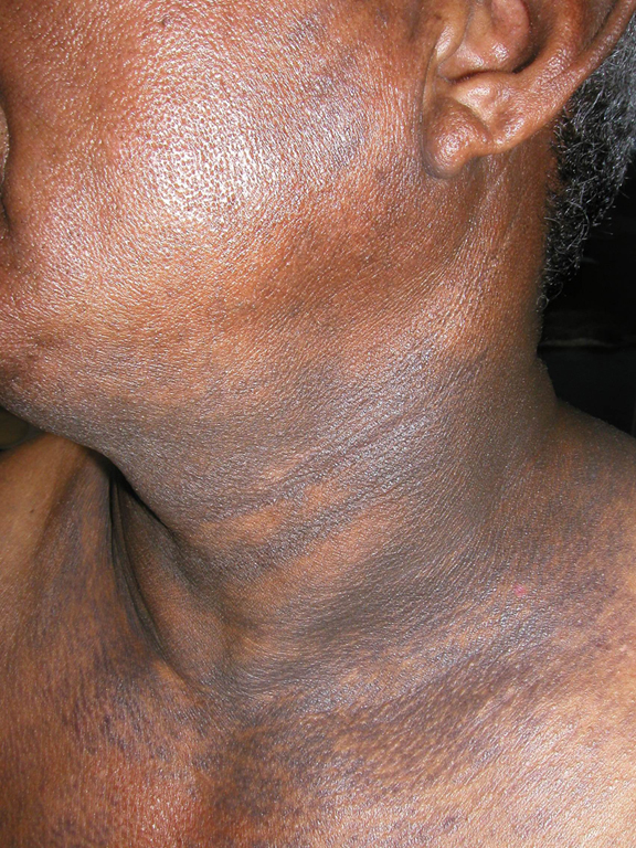
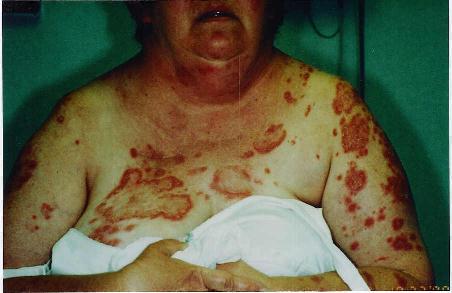
EAC presents as one or more lesions that begin as erythematous or urticarial-like papules and enlarge by peripheral extension to form ringed arcuate, or polycyclic figures. These spread gradually to form large rings with central clearing, the edges of the lesions often advancing several millimeters a day. Dull red, brown, or violet-tinged centers are left behind the advancing margins. Two subgroups have been described: deep and superficial. The superficial variant differs only by the presence of a characteristic trailing scale, a delicate annular rim
of scale that trails behind the advancing edge of erythema. Itching is variable but seldom intense. The common sites of involvement are the buttocks, thighs, and upper extremities, but any areas may be involved. Rarely, if ever, are the palms, soles, scalp, or mucous membranes involved. The disease may persist with periodic fluctuations over many years. There is no residual scarring as lesions resolve.
In the classic deep form, the histologic features are a superficial and deep perivascular lymphocytic infiltrate characterized by a tightly cuffed “coat sleeve-like” pattern present in the middle and lower dermis, with a usually unremarkable epidermis. This corresponds to the greater induration and elevation than is seen in the superficial form of EAC. In the superficial variant, the perivascular tightly cuffed lymphohistiocytic infiltrate is more mild and superficial, with endothelial cell swelling and focal extravasation of erythrocytes in the papillary dermis. In addition, there is focal epidermal spongiosis and focal parakeratosis. These findings correspond to the characteristic trailing scale. The central area of clearing may contain dermal melanophages.
TREATMENT:
Initially, the rash resolved with oral terbinafine (Lamisil) and topical halobetasol propionate (Ultravate) cream, but then recurred again in April 1998. At that time the patent was started on topical steroids and antipruritic medications for symptomatic relief, with significant improvement. She flared again in August 1998; topicals were resumed and terbinafine restarted. After 3 weeks of no improvement, she began a 3-week taper of oral prednisone with significant improvement. The patient’s condition then waxed and waned over the next several months with significant improvement only to oral prednisone. Then only after receiving X-ray therapy to the 1.5 cm right pulmonary nodule in December 1998, the lesions gradually began clearing. The pulmonary nodule also decreased in size to less than 0.5 cm. Fourteen months after receiving the X-ray therapy, the patient remained clear.
A comprehensive search for the underlying cause is the primary goal of treatment. The skin lesions will usually resolve once the underlying cause has been treated successfully. Systemic corticosteroids usually suppress EAC, but recurrence is common on discontinuation, as was in our case. Topical steroids may be helpful when there is marked epidermal involvement. Empirical use of antibiotics, antifungal, or anticandidal agents has sometimes been useful. Initially, our case responded to oral antifungal agents but later recurred. Sedating antihistamines can be employed for associated pruritus. The use of calcipotriol (Dovonex) was reported in the case of a 73-year-old female with a three-year history of EAC resistant to topical and systemic glucocorticoids, antifungals, and PUVA treatment. After three months of treatment with topical calcipotriol, the lesions cleared completely and did not recur during a six month follow-up period.
REFERENCES:
Bolognia, J. L., et al. (2003). Dermatology (1st ed., pp. 303–306). Mosby.
Mahood, J. M. (1983). Erythema annulare centrifugum: A review of 24 cases with special reference to its associations with underlying disease. Clinical and Experimental Dermatology, 8, 383. [PMID: 6629898]
Bressler, G. S., & Jones, R. E. (1981). Erythema annulare centrifugum. Journal of the American Academy of Dermatology, 4, 597–602. [PMID: 7031345]
Gniadecki, R. (2002). Calcipotriol for erythema annulare centrifugum. British Journal of Dermatology, 146(2), 317–319. [PMID: 11896866]