Presenter: Valerie Johnson, D.O., Marya Cassandra, D.O., Greg Houck, D.O., Kristin Witfill, D.O., Matt Muellenhoff, D.O., Thi Tran, D.O.
Dermatology Program: Sun Coast Hospital (NOVA Southeastern University)
Program Director: Richard Miller, D.O.
Submitted on: March 30, 2004
CHIEF COMPLAINT: Recurring and rapidly enlarging nodule on the right middle finger
CLINICAL HISTORY: A 7 month-old, healthy appearing, well-nourished male presented with a recurring and rapidly enlarging nodule on the right middle finger that parents stated was cosmetically disfiguring. The nodule had previously been excised by a hand surgeon when the patient was 2 months old. The mother was concerned about the recurrence and prognosis.
PHYSICAL EXAM:
0.5cm firm, yellow-pink, the dome-shaped nodule was noted on the right, lateral, distal third digit. An adjacent, well-healed scar from the previous surgery was also present. There was no evidence of any physical impairment secondary to the growth.

LABORATORY TESTS: N/A
DERMATOHISTOPATHOLOGY:
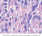
This is a non-encapsulated tumor composed of spindle-shaped myofibroblasts intermixed with collagen bundles. It may extend from the epidermis down into the subcutaneous tissue. Myofibroblasts have pathognomic eosinophilic, cytoplasmic inclusion bodies, which stain red with Masson’s Trichrome, deep purple with phosphotungstic acid hematoxylin (PTAH). These inclusions are mostly aggregates of actin. (no histopathology available from our case, therefore, an example is taken from Bolognia)

DIFFERENTIAL DIAGNOSIS:
1. Supernumerary digit
2. Acral angiofibroma
3. Acral fibrokeratoma
4. Infantile digital fibroma


