Presenter: Charmaine Jensen DO, Rene Bermudez DO, Dimitry Palceski DO, Theresa Ng DO
Dermatology Program: Cuyahoga Falls General Hospital
Program Director: Schield Wikas DO
Submitted on: April 21, 2004
CHIEF COMPLAINT: “Raised bumps all over my body”
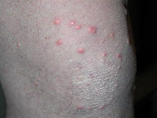
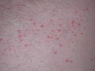
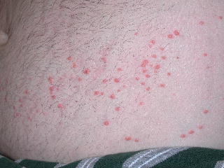
CLINICAL HISTORY: A 40-year-old 120kg Caucasian male presented to our clinic with multiple, moderately pruritic yellow hard papules on his knees, thighs, buttocks, back, abdomen, and elbows. He had been in a hot tub two weeks prior to the onset of his lesions. The lesions appeared suddenly on his knees and elbows and subsequently spread to his abdomen, lower back, thighs, and buttocks. He presented to the emergency department and was diagnosed with molluscum contagiosum. He has treated these lesions with Retin-A cream and cryosurgery. No other family members are affected. He has a history of diabetes mellitus type 2 and had not been on his medications for several months because of financial constraints. He denied having any nausea, vomiting, or abdominal pain.
PHYSICAL EXAM:
The patient is a 40-year-old 120kg Caucasian male with multiple 2-4mm yellow hard papules on his knees, thighs, buttocks, back, abdomen, and elbows. Some have an erythematous halo in which the patient attributed to the previous cryosurgery. The lesions did not exhibit any central umbilication or Koebner phenomenon. There was no evidence of Grey Turner’s sign or Cullen’s sign.
LABORATORY TESTS:
Glucose 339 mg/dL
Hemoglobin A1C 13.1%
Cholesterol 4,990 mg/dL
Triglycerides >5,750 mg/dL
HDL 12 mg/dL
LDL *
VLDL *
Blood was grossly lipemic.
*Calculations for LDL and VLDL are not accurate when triglycerides are >400.
DERMATOHISTOPATHOLOGY:
The epidermis is unremarkable. Foamy histiocytes are noted throughout the reticular dermis. There is a mixed inflammatory infiltrate present containing neutrophils and lymphocytes. Extracellular lipid depositions that are seen as artifactual clefts filled with a wispy faint blue-gray material are also present in the dermis.

DIFFERENTIAL DIAGNOSIS:
- Eruptive histiocytomas
- Granuloma annulare
- Juvenile xanthogranulomas
- Eruptive xanthomas
- Molluscum contagiosum


