CORRECT DIAGNOSIS:
Infantile Hemangiomas
DISCUSSION:
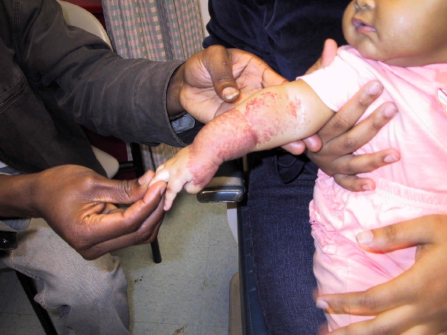
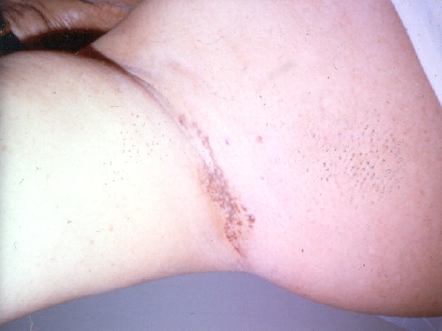
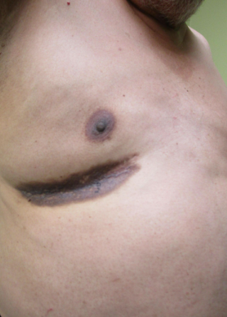
Infantile hemangiomas manifest at birth in 30% of patients. It is the most common and benign tumor of infancy. Other patients may manifest at 2 weeks to 2 months of age with a pale macule. Usually, 1 – 60 mm in diameter hemangiomas are found on the head and neck (60% of cases) though may present anywhere on the body. Rarely an entire extremity is involved. Clinically the lesion appears as a dull red dome-shaped lesion, with sharp borders and is easily compressible. When involution occurs one may see streaks or islands of white. Hemangiomas tend to grow over the first year of life then stabilize before involuting. 50% will involute by 5 years of age and 70% by 7 years of age. Depending on the size and duration of the hemangioma the patient may be left with atrophy, telangiectasias, and an anetoderma type redundancy.
The malformation is associated with 7% of hemangiomas and may include such syndromes as PHACES and Kippel-Trenaunay Syndrome. PHACES is associated with posterior fossa malformations, hemangiomas, arterial malformations, coarctation of the aorta, and eye abnormalities. Kippel-Trenaunay Syndrome has a triad of nevus flammeous (confined to skin extremity), varicose veins (venous malformations), and soft tissue hypertrophy of the affected limb.
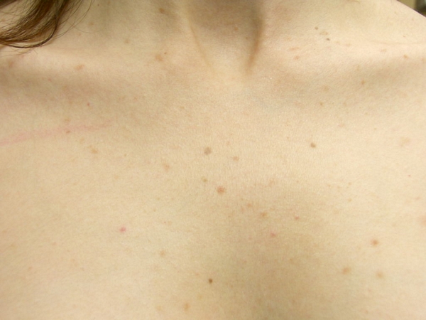
Multiple Hemangiomas, ranging in size from 1-10 mm, are referred to as benign neonatal hemangiomatosis. Diffuse Neonatal Hemangiomatosis has a poor prognosis and involves the viscera including CNS, lung, and liver. These lesions can lead to complications including GI or CNS bleeding, and respiratory failure. Histologically hemangiomas resemble primitive endothelial cells though they lack Weibel-Palade bodies. Crystalloid inclusions typical of embryonic endothelium are appreciated. There is a sequence of events that may be appreciated as the intraluminal proliferates, flattens and the lumen becomes more apparent. Once fibrosis begins the lumen shrinks and involution progresses.
TREATMENT:
Hemangiomas should be treated aggressively if they hemorrhage, ulcerate, thrombose, compromise airways, or vision. Early intervention for cosmetic results is important especially for school-age children with lesions on the face. Cryotherapy or laser ablation of early lesions is unaffected. Pulse dye lasers (PDL) helps with residual telangiectasia. Surgery is an excellent option for small pedunculated lesions or patients with the Cyrano defect. Cyrano defects are those hemangiomas at the tip of the nose that has become bulbous. In addition, compressive wraps may improve the outcome of hemangiomas on an extremity.
Treatment if not surgical may include intralesional steroids or oral prednisone. Hemangiomas stabilize (stop growing) in 30% of patients on 2-3mg/kg/day of PO prednisone within 3-21 days. Ulcerated lesions take 2 weeks to respond. Lesions go on to involute at approximately 30-90 days of PO treatment. Repeat courses may be required with rebound growth. Maintenance with low dose prednisone for 1 year may prevent rebound. 30% of patients don’t respond to PO prednisone. Recombinant interferon-alpha 2a or 2b has been successful in 80% of patients on 1-3 million units/m2/day. Responses may take 6-10 months. Side effects include thyroid dysfunction and neurotoxicity (spastic diplegia).
REFERENCES:
Odom, R. B., James, W. D., & Berger, T. G. (2000). Andrews’ diseases of the skin: Clinical dermatology (9th ed., pp. 740, 750-751). W. B. Saunders Company.


