CORRECT DIAGNOSIS:
Zoon’s balanitis
DISCUSSION:
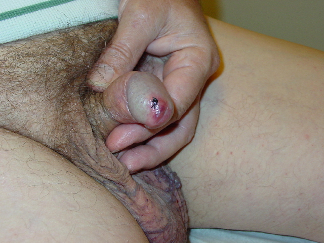
Zoon’s Balanitis is a condition found on the glans penis and/or inner surface of the prepuce of the uncircumcised, middle-aged to the older male. We report a case of a 58-year-old male who presented to our clinic with a persistent, glistening plaque on the penis of 5 years duration. The differential diagnosis is quite vast, including erythroplasia of Queyrat, thus biopsy is essential to rule out underlying neoplasms and infections.
Zoon’s balanitis goes by many names, including plasma cell balanitis, balanitis circumscripta plasmacellularis, and plasma cell mucositis. The female equivalent is termed plasma cell vulvitis. The oral mucosal equivalent is termed plasma cell orificial mucositis. Clinically, Zoon’s balanitis presents most often as a solitary, glistening, red or cayenne pepper-colored, persistent plaque on the glans penis or inner surface of the prepuce of the uncircumcised male. The etiology is unknown, however, it occurs almost exclusively in the uncircumcised male. It has been pointed out that plasma cells frequently predominate in the inflammatory response at mucocutaneous junctions in a variety of benign and malignant processes.
The lesion itself is usually asymptomatic aside from some mild pruritus. Patients may also complain of irritation with intercourse. Further, secondary infections with candidal organisms are not uncommon. The area raises a concern of malignancy by the patient and physician alike. The differential diagnosis for balanoposthitis is large including Candida albicans, Groups A and B streptococcus, Gonococcus, Genital herpes, Gardnerella vaginalis, Trichomonas vaginalis, Mycoplasma, Chlamydia trachomatis, Syphilis, nonspecific intertrigo, traumatic lesions, Zoon’s balanitis, psoriasis vulgaris, Reiter’s syndrome, allergic contact dermatitis, fixed drug eruption, drug-induced erosions, squamous cell carcinoma, and Extramammary Paget’s disease. Diagnosis is a histologic one. Often, topical corticosteroids and antifungals are used empirically without success. This prompts biopsy for a more definitive diagnosis, specifically to rule out neoplasia.
Histologically, the epidermis appears thinned, often showing an absence of the upper layers. If present, the epidermis is often distinctive in that in addition to being thinned and flattened, it is composed of diamond-shaped, pancake-like keratinocytes that are separated by uniform intercellular edema. It is not uncommon to have erythrocytes percolating up through the epidermis. The upper dermis demonstrates a lichenoid infiltrate with copious plasma cells. In some cases, the number of plasma cells is low. Capillary dilatation is also not uncommon.
TREATMENT:
The patient was counseled extensively on this condition. Topical therapy failed to provide relief of symptoms. He was referred to urology for circumcision.
Treatments start with topical therapies. Mild topical corticosteroids are the initial treatment of choice, however, recurrence upon their discontinuation is the rule. Circumcision is curative in nearly all cases. Close follow-up is recommended.
REFERENCES:
Odom, R. B., James, W. D., & Berger, T. G. (2000). Andrews’ diseases of the skin: Clinical dermatology (9th ed., pp. 840-842). W. B. Saunders Company.
Baldwin, H. E., et al. (1989). The treatment of Zoon’s balanitis with the carbon dioxide laser. Journal of Dermatologic Surgery and Oncology, 15, 491.
Brodin, M. (1980). Balanitis circumscripta plasmacellularis. Journal of the American Academy of Dermatology, 2, 33.
Ferrandiz, C., & Ribera, A. (1984). Zoon’s balanitis treated by circumcision. Journal of Dermatologic Surgery and Oncology, 10, 622.
Fitzpatrick, T. B., et al. (1999). Dermatology in general medicine (5th ed., pp. 1350-1351). McGraw-Hill.
Elder, D., et al. (1997). Lever’s histopathology of the skin (8th ed., pp. 710-711). Lippincott-Raven.