CORRECT DIAGNOSIS:
X-linked ichthyosis
DISCUSSION:
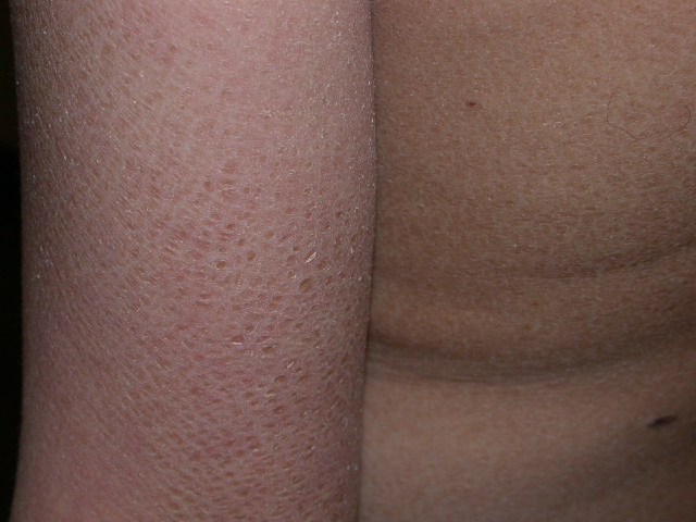
X-linked ichthyosis is the second most common type of ichthyosis with ichthyosis vulgaris being most common. It affects between 1 in 2,000 to 6,000 males and has no geographic or racial predilections. It typically develops at birth or early infancy usually between 2 and 6 weeks of age with light-colored scaling with progression to large, dark scales giving the skin a dirty appearance. The scales are prominent on the extensor surfaces, pre-auricular, neck, and upper trunk with or without flexural involvement. The palms and soles are typically spared and hair and nails are normal.
Extracutaneous manifestations are possible including comma-shaped corneal opacities in affected males and female carriers. These are asymptomatic and do not affect visual acuity. Affected patients have an increased risk of cryptorchidism and testicular cancer even without a history of undescended testes. Placental steroid sulfatase deficiency is associated with prolonged labor and failure to progress during the birth of the affected patient. Since steroid sulfatase normally hydrolyzes cholesterol sulfate and sulfated steroid hormones, a deficiency leads to low estrogen levels resulting in difficulty initiating labor and a higher number of women requiring Cesarean sections.
X-linked ichthyosis is due to a genetic disorder of keratinization leading to retention ichthyosis. It is caused by a deficiency of steroid sulfatase enzyme resulting from abnormalities in its coding gene. The gene is mapped to the distal part of the short arm of the X22 chromosome. This deficiency leads to the accumulation of cholesterol sulfate and decreased cholesterol in the stratum corneum which causes persistent keratinocyte adhesion and abnormal desquamation.
The dermatopathology is nonspecific but generally shows mild to moderate compact orthokeratosis, a normal to thickened granular layer, and mild to moderate acanthosis.
The differential diagnosis includes other common ichthyoses. The distinction is made clinically and with the aid of histopathology. Ichthyosis vulgaris is the most common type of ichthyosis and shows fine, white scales over the extensors with flexural sparing, keratosis pilaris, and palmoplantar involvement. A decreased or absent granular layer is seen. Lamellar ichthyosis and congenital ichthyosiform erythroderma both present as colloidion babies at birth. Later, lamellar ichthyosis shows large quadrilateral plate-like scales and palmoplantar involvement with a normal granular layer seen on histopathology. Congenital ichthyosiform erythroderma shows finer white scales over the body, erythroderma, and alopecia as well as a normal to decreased granular layer on histopathology. Bullous ichthyosiform erythroderma shows erythroderma, scaling, and bullae early on which progress to intertriginous dark scales over time. On histopathology, an increased granular layer with vacuolization of the granular and spinous layers is seen.
Diagnosis is made by demonstrating gene deletion, lack of enzyme activity, or an increase in one of the substrates of steroid sulfatase. Fluorescence in situ hybridization (FISH) study is performed on metaphase chromosomes from peripheral blood cells using a steroid sulfatase probe. The absence of a hybridization signal indicates a microdeletion of the steroid sulfatase gene. A steroid sulfatase assay will show reduced or absent enzyme activity in leukocytes, skin fibroblasts, and keratinocytes. This is used to detect carriers and affected patients. Elevated cholesterol sulfate levels in the serum detected by chromatography or spectrophotometry may be used as well as PCR and lipoprotein electrophoresis since increased cholesterol sulfate increases electrophoretic mobility of low-density lipoproteins. Maternal urine can be used to detect high levels of sulfated estrogen precursors or low levels of estriol and amniotic fluid analysis may be used prenatally to measure placental sulfatase activity and elevated DHEAS.
TREATMENT:
Our patient was treated with Lac-hydrin 12% cream BID. Topical keratolytics such as ammonium lactate which contains lactic acid, an alpha-hydroxy acid with keratolytic action via disadhesion of corneocytes is the most commonly used and successful treatment although emollients, hydrating agents and topical retinoids may be tried. Oral retinoids are reserved for rare or more significantly affected patients.
REFERENCES:
Ellison, P., et al. (1996). Chronic dark-brown scales. Archives of Dermatology, 23, 594-597.
Hernandez-Martin, A., et al. (1999). X-linked ichthyosis: An update. British Journal of Dermatology, 141, 617-627.
Hirato, K., et al. (1991). Steroid sulfatase activities in human leukocytes: Biochemical and clinical aspects. Endocrinology Japan, 38(6), 597-602.
Nomura, K., et al. (1995). A study of the steroid sulfatase gene in families. Acta Dermato-Venereologica, 75(5), 340-342.
Paller, A. S. (1994). Laboratory tests for ichthyosis. Dermatologic Clinics, 12(1), 99-107.


