CORRECT DIAGNOSIS:
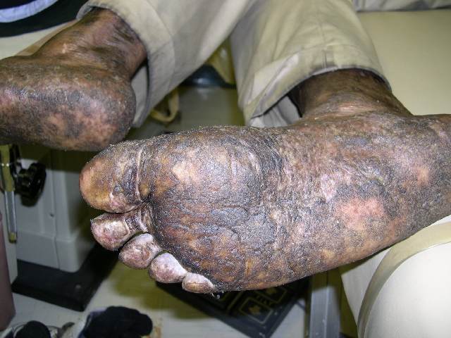
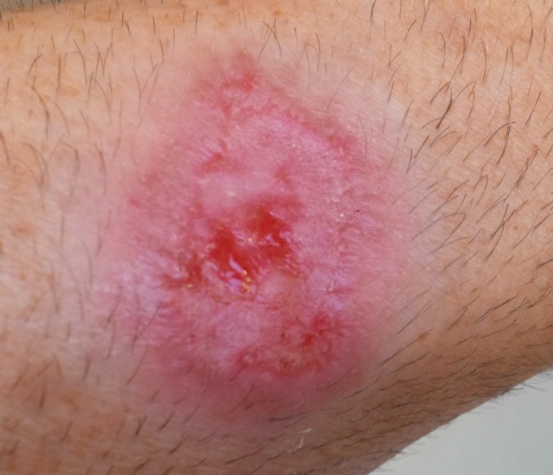
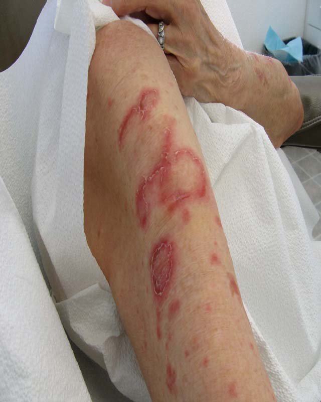
Mycosis fungoides
DISCUSSION:
Mycosis fungoides is the most common type of cutaneous T-cell lymphoma (CTCL) as it accounts for approximately 50% of the cases. There is an incidence of 0.3 per 100,000 individuals. Median age of diagnosis is 55-60, and males are more commonly affected. Patients generally progress from patches (premycotic stage) to plaques and finally nodules. The disease is usually located in the “bathing trunk” distribution. Prior to proper diagnosis, patients initially present with a nonspecific eczematous or psoriasiform dermatitis with inconclusive skin biopsies.
The distinguishing histologic features include nests of atypical cells with highly convoluted and hyperchromatic nuclei (Pautrier’s microabscesses) and epidermotropism of lymphocytes. Immunophenotyping typically stains positive for CD3, CD4, and CD45 RO and mostly negative for CD8. Unique to our case was the CD30+ large cell anaplastic transformation, which is associated with a poor prognosis.
Our patient presented with classic leonine faces. The differential that should be considered is as follows: carcinoid, chronic actinic dermatitis, cutis verticis gyrata, leishmaniasis, leprosy, lipoid proteinosis, lymphma/leukemia, MF, multicentric reticulohistiocytosis, multiple keratoacanthoma syndrome, progressive nodular histiocytoma, sarcoidosis, and scleromyxedema.
Prognosis is dependent on the stage, extent of skin lesions, and presence of extracutaneous manifestations. Patients with limited patch/plaque stage have normal life expectancy. Lymph node and visceral involvement, as well as transformation into large T-cell lymphoma portends an aggressive course.
TREATMENT:
Our patient was placed on topical corticosteroids and referred to an oncologist for systemic workup.
Treatment options include topical corticosteriods, topical chemotherapy (including nitrogen mustard and carmustine), radiotherapy, phototherapy, systemic chemotherapy (CHOP), biologic response modifiers, interferons, and retinoids. Novel immunomodulatory therapies such as receptor-targeted cytotoxic fusion proteins and various vaccines have been used.
REFERENCES:
Arnold, A., Odom, R. B., & James, W. D. (2000). Andrews’ diseases of the skin. Philadelphia, PA: Elsevier.
Bolognia, J. L., Jorizzo, J. L., Rapini, R. P., et al. (2003). Dermatology. Spain: Mosby.
Paulli, M., & Berti, E. (2004). Cutaneous T-cell lymphomas (including rare subtypes): Current concepts. Haematologica, 89(11), 1372-1379.


