Presenter: Matthew Smetanick, D.O., Gregg Severs, D.O., J. Greg Brady, D.O.
Dermatology Program: Frankford Hospital/PCOM
Program Director: Stephen M. Purcell, D.O.
Submitted on: September 28, 2006
CHIEF COMPLAINT: Painful, swollen left foot
CLINICAL HISTORY: A 69-year-old, otherwise healthy woman was seen in our office for a painful, swollen left foot. She reported a two-week history of worsening symptoms after stepping on a sharp object while walking barefoot at home. The patient experienced pain at the base of her left first toe, but could not identify an obvious puncture wound or foreign body at the time of injury. Initially, the patient was seen at an urgent care center and was treated with prednisone for a suspected bite reaction on her toe. The patient was then seen five days later by her primary care physician and received cephalexin for suspected cellulitis. Her prednisone was also discontinued at that time.
Her past medical history was significant for hypertension and hyperlipidemia. Medications included olmesartan, rosuvastatin, and a multivitamin. The patient admitted to having chills a few days prior to her presentation at our office. The patient was sent to the emergency room for cultures and radiographic studies. She was subsequently admitted and placed on empiric antibiotic therapy with clindamycin and vancomycin. The patient was also started on itraconazole to cover a possible fungal infection, pending tissue biopsy, and culture results.
PHYSICAL EXAM:
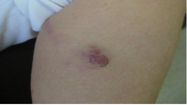
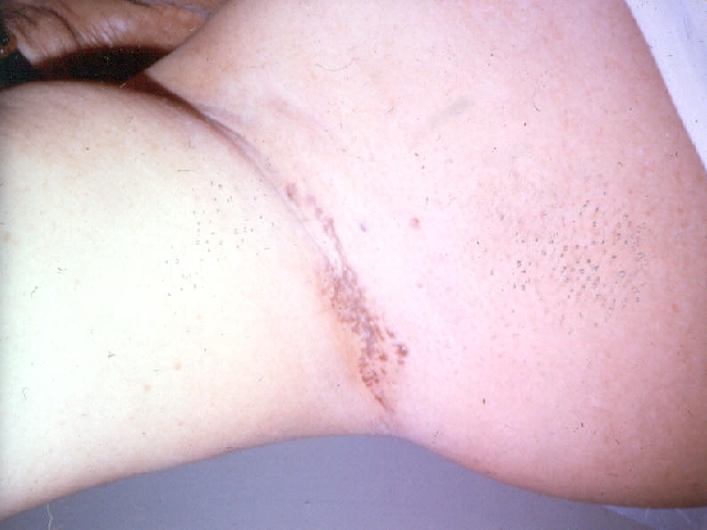
Initial examination revealed erythema, swelling, and increased color of the dorsal left foot with erythematous, lymphangitic streaking extending from the left lower medial leg up to the groin. Approximately two days later, the patient developed tense, suppurative bullae extending superiorly in a sporotrichoid pattern up her left medial leg and left posterior thigh. The lesions were drained, debrided, and biopsied by dermatology.


Granules were not grossly identified while performing these procedures. An x-ray and MRI of the left foot demonstrated no evidence of osteomyelitis but did reveal extensive subcutaneous edema consistent with cellulitis.
LABORATORY TESTS:
Punch biopsy specimens were sent for tissue culture, which eventually yielded gram-positive, branching filaments, consistent with Nocardia species. Final culture results subsequently isolated Nocardia braziliensis as the causative agent.
DERMATOHISTOPATHOLOGY:
A punch biopsy specimen from one of the debrided areas of the left leg revealed spongiotic changes of the epidermis, superficial perivascular and interstitial inflammation with neutrophils and deep dermal and subcutaneous neutrophilic abscess formation. No asteroid bodies or any evidence of the Splendore-Hoeppli phenomenon were identified. No microorganisms were identified by hematoxylin and eosin sections. PAS, GMS, AFB, and Gram stains also revealed no definitive fungal, mycobacterial, or bacterial organisms.
DIFFERENTIAL DIAGNOSIS:
1. Lymphocutaneous Nocardiosis
2. Sporotrichosis
3. Cutaneous tuberculosis
4. Other mycobacterium