CORRECT DIAGNOSIS:
Lymphocutaneous Nocardiosis caused by Nocardia brasiliensis
DISCUSSION:
Nocardia species are gram-positive, aerobic, weakly acid-fast bacilli with fine branching filaments. Colonies will grow on Löwenstein-Jensen culture media, Sabouraud’s glucose agar, and blood agar and are usually rough with a velvety surface. They typically have a white or light orange color and a characteristic moldy or earthy odor. Optimum growth occurs at 37º, but growth is slow. Colonies may not be visible for 48 to 72 hours of incubation, and all culture specimens of possible Nocardia should be observed for a full two weeks. Histopathology shows a mixed acute abscess and granulomatous response with fibrosis. Occasionally, the bacteria are clumped together with surrounding homogeneous eosinophilic material. This is known as the Splendore-Hoeppli phenomenon. More often, the bacteria are loosely dispersed and poorly demonstrated with hematoxylin-eosin stain. They are Gram-positive, Grocott-silver-positive, and weakly acid-fast with modified Ziehl-Neelsen stain.
Nocardia species are native to the soil and decaying vegetable matter and only accidentally infect man. Infection occurs after direct inoculation of the skin or by inhalation. The Nocardia species pathogenic in man include N. asteroides, N. brasiliensis, and N. otitidiscaviarum. In North America, most Nocardia infections are caused by N. asteroides, whereas in Latin America, most are caused by N. brasiliensis. N. asteroides usually presents as pleuropulmonary or less commonly skin infection in immunosuppressed hosts, whereas N. brasiliensis more commonly causes primary cutaneous disease. In laboratory animals, the virulence of N. brasiliensis exceeds that of N. asteroides, which supports observations that N. asteroides is predominantly an opportunistic pathogen, whereas N. brasiliensis causes skin infections in normal hosts.
Nocardiosis is divided into systemic and cutaneous types. Systemic nocardiosis almost exclusively begins in the respiratory tract. Cutaneous nocardiosis can have one of four clinical manifestations: (1) mycetoma, (2) lymphocutaneous (sporotrichoid) infection, (3) superficial skin infections such as cellulitis, abscesses, ulcers, or granulomas, and (4) disseminated disease with cutaneous involvement.
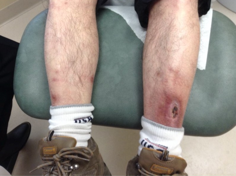
As seen in our case, the lymphocutaneous (sporotrichoid) infection of nocardiosis usually begins as an ulcerated papule at the site of inoculation, followed by advancing lymphangitis and subcutaneous erythematous nodules along the lymphatic drainage. Typical lesions are located on the extremities with a history of a puncture wound or farm/garden work. Most patients lack systemic manifestations such as fever, leukocytosis, or weight loss.
Many cases of cutaneous nocardiosis mimic more familiar skin eruptions, such as cellulitis or bacterial abscesses, and are treated with drainage and antibiotics without Gram stain or culture. Other causes for the sporotrichoid pattern of spread besides N. brasiliensis and N. asteroides, includes Sporothrix schenckii, Coccidioides immitis, Blastomyces dermatitidis, Histoplasma capsulatum, tularemia, lymphatic tumors, Mycobacterium marinum, Mycobacterium kansasii, and Mycobacterium chelonei. Potential clues to the diagnosis of Nocardia infection include history of traumatic inoculation, occupational exposure (gardening, farming), the tendency for cutaneous infections to recur, worsening of infection despite “standard” antibiotic treatment, chronic cutaneous infections with “negative cultures” (see culture media and duration requirements above), acid-fast organisms, and sulfur granules.
Nocardiosis often requires months of antibiotic therapy. The drugs of choice are sulfonamides, while other reports suggest efficacy with minocycline and aminoglycosides, especially amikacin. Surgical drainage of suppurative cutaneous infections is also an important part of treatment.
TREATMENT:
The patient’s antibiotic regimen was changed to trimethoprim/sulfamethoxazole for appropriate coverage. Further debridement and abscess drainage was also required in our office.
Left medial leg. Demonstrating lesions two weeks after discharge from hospital:

Left medial leg. Two weeks after hospital discharge, demonstrating healthy, granulation tissue following further debridement in our office:

She is currently following with infectious disease and dermatology. Her wounds have been healing successfully, and the patient must remain on oral trimethoprim/sulfamethoxazole for the next few months before a complete resolution is expected.
REFERENCES:
Kalb, R. E., Kaplan, M. H., & Grossman, M. E. (1985). Cutaneous nocardiosis: Case reports and review. Journal of the American Academy of Dermatology, 13, 125-133. PMID: 4014286
Karakayali, G., Karaarslan, A., Artuz, F., Alli, N., & Tereli, A. (1998). Primary cutaneous Nocardia asteroides. British Journal of Dermatology, 139, 919-920. PMID: 9885603
Aly, R., & Maibach, H. I. (1999). Subcutaneous mycoses: Mycetoma, chromoblastomycoses, sporotrichosis. In Atlas of infections of the skin (pp. 67-70). New York: Churchill Livingstone.
Elder, D., Elenitsas, R., Jaworsky, C., & Johnson, B. (1997). Bacterial disease. In Lever’s histopathology of the skin (pp. 496-497). Philadelphia: Lippincott Williams & Wilkins.


