CORRECT DIAGNOSIS:
Porokeratosis of Mibelli
DISCUSSION:
Porokeratosis is a disorder of keratinization characterized histologically by the presence of a cornoid lamella. It was first described by Mibelli in 1893. Today, five clinical variants exist: (1) classic porokeratosis of Mibelli, (2) disseminated superficial (DSP) and disseminated superficial actinic porokeratosis (DSAP), (3) porokeratosis palmaris et plantaris disseminata (PPPD), (4) linear, and (5) punctate porokeratosis.
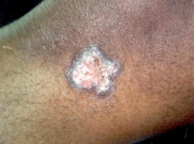
Clinically, lesions start as small, brownish, keratotic papules, which slowly enlarge forming irregular, annular plaques with a well-demarcated, raised hyperkeratotic border. The center of the lesion is usually atrophic. Lesions can range in size from a few millimeters to several centimeters. Usually, only a few lesions are present and they can present anywhere. However, they are typically found on acral areas of the extremities, thighs, and perigenital region. Lesions can spread by koebnerization.
The onset of porokeratosis of Mibelli is during childhood and males are more often affected than females. Familial studies suggest an autosomal dominant mode of inheritance. Lesions usually increase in size and number over time and tend to be asymptomatic.
Histologically, porokeratosis of Mibelli is characterized by a prominent cornoid lamella- a thin column of parakeratotic cells that extend through the surrounding orthokeratotic stratum corneum. Other features include an absent or decreased granular zone beneath the column of parakeratosis, vacuolated and/or dyskeratotic cells in the spinous layer, epidermal hyperplasia, and dilated capillaries associated with a moderately dense lymphohistiocytic infiltrate in the papillary dermis.
The etiology of porokeratosis is unknown. To some investigators, the cornoid lamella represents a defect in cornification. Others proposed that porokeratosis of Mibelli could be related to a mutant cellular clone of epithelial cells in the epidermis, and that the cornoid lamella represents the boundary between the abnormal clonal population and the normal epithelium.
Acquired immune deficiency is frequently found as a prelude to the development of porokeratosis and can account for up to 50% of new cases. Specifically, the reported incidence of porokeratosis occurring after organ transplant ranges from 0.34%-11%. The majority of patients developed disseminated superficial porokeratosis.
Although the pathogenesis of PK is unknown, it was for many years viewed as a benign entity. However, several cases of malignancy arising in lesions of porokeratosis were reported. The potential for malignancy has not been well defined and the risk of malignant transformation is unknown. However, authors have reported malignant degeneration in ~10%. The most commonly reported malignant lesions include Bowen’s disease, SCC, and rarely BCC. Malignant lesions have been reported to arise in larger, extremity lesions. Older patients and those with the longstanding disease are at increased risk.
In spite of a poorly defined pathogenesis, porokeratosis displays a potential for malignancy and, at the minimum, careful observation is warranted.
TREATMENT:
For this patient, smaller lesions were treated via cryotherapy with liquid nitrogen. Larger lesions were initially treated with Tazarotene 0.1% cream bid. However, topical treatment with 5-FU is planned if lesions are unresponsive to tazarotene.
Other treatment options include excision, cryotherapy, electrodesiccation, dermabrasion, CO2 laser, 585 PDL, keratolytics, topical 5-FU, topical 5% imiquimod, topical diclofenac sodium, oral and topical retinoids. Patients should also avoid radiation, immunosuppression, and excessive UV exposure.
REFERENCES:
Kanitakis, J., et al. (2001). Porokeratosis in organ transplant recipients. Journal of the American Academy of Dermatology, 44(1), 1-4. PMID: 11136927
Raychaudhuri, S. P., & Smoller, B. R. (1992). Porokeratosis in immunosuppressed and nonimmunosuppressed patients. International Journal of Dermatology, 31, 781-782. PMID: 1449938
Reed, R. J., & Leone, P. (1970). Porokeratosis: A mutant clonal keratosis of the epidermis. I. Histogenesis. Archives of Dermatology, 101, 340-344. PMID: 4919032
Rio, B., et al. (1997). Disseminated superficial porokeratosis after autologous bone marrow transplant. Bone Marrow Transplantation, 19, 77-79. PMID: 9033651
Sasson, M., & Krain, A. D. (1996). Porokeratosis and cutaneous malignancy: A review. Dermatologic Surgery, 22, 339-342. PMID: 8727465
Wade, T. R., & Ackerman, A. B. (1980). Cornoid lamellation: A histologic reaction pattern. American Journal of Dermatopathology, 2, 5-15. PMID: 6100745


