Presenter: Alice Do, DO, Brian Kopitzki, DO, Chris Buatti, DO
Dermatology Program: Genesys / Michigan State University
Program Director: Kimball Silverton, DO
Submitted on: February 18, 2008
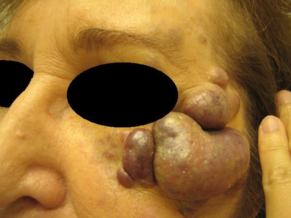
CHIEF COMPLAINT: Facial mass.
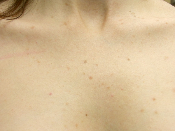
CLINICAL HISTORY: A 73-year-old Caucasian woman presented with a 20-year history of violaceous masses of the left periocular area and left chest that has waxed and waned. These lesions were asymptomatic. 10 years ago, the lesions were biopsied and diagnosed as a low-grade B cell lymphoma without systemic involvement, and no chemotherapy was indicated at that time. Over the years, the lesions continued to wax and wane, but recently, the lesions have gotten larger.
PHYSICAL EXAM:
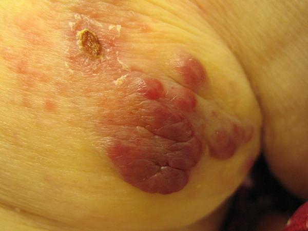
There were large, violaceous nodules in the left peri-ocular area. There were also violaceous nodules, plaques, and patches on the left anterior chest and right posterior flank. No lesions were noted on the legs or lower body. There was no fever, lymphadenopathy, or hepatosplenomegaly.
LABORATORY TESTS:
CBC and chemistries (including LFT’s and Cr) were normal. A peripheral blood smear was normal. Bone marrow aspirate and biopsy were normal. Clonality studies revealed a lambda restriction. A PET scan showed uptake in the left periorbital subcutaneous tissue, left upper anterior chest wall, & right axilla. These areas corresponded to cutaneous lesions.
DERMATOHISTOPATHOLOGY:
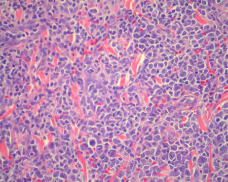
The H & E biopsy shows an expansion of the dermis by large atypical lymphoid cells with hyperchromatic nuclei, prominent nucleoli, and mitotic figures. These cells are growing in a diffuse pattern, and no follicular centers are seen. A grenz zone separates these lesional cells from the epidermis, which shows a lack of epidermotropism.
Immunostains revealed the following: LCA+, CD20+, CD79a+, BCL2+, Vimentin+. CD10-, CD30-, CD45RO-, Factor VIII-, CK20-, Synaptophysin-, Pancytokeratin-, CD34-. Peripheral CD3+, CD5+, and CD43+ represented reactive T cells to the lesional neoplastic cells.
Diffuse large B-cell non-Hodgkin’s lymphoma, high grade.
DIFFERENTIAL DIAGNOSIS:
1. Lymphangioma
2. Hodgkin’s lymphoma
3. Non- Hodgkin’s lymphoma
4. CTCL
5. CD30+ Anaplastic Large Cell Lymphoma


