Presenter: Patrick Keehan, D.O.
Dermatology Program: K.C.O.M. – Texas Division
Program Director: Bill V. Way, D.O.
Submitted on: April 2, 2008
CHIEF COMPLAINT: Asymptomatic lesion on the back x 8 years
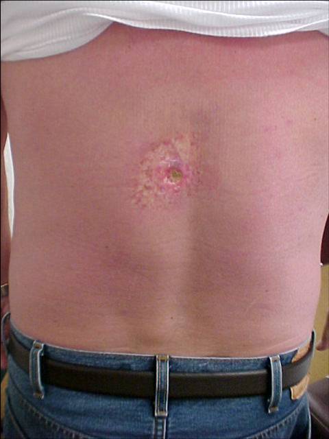
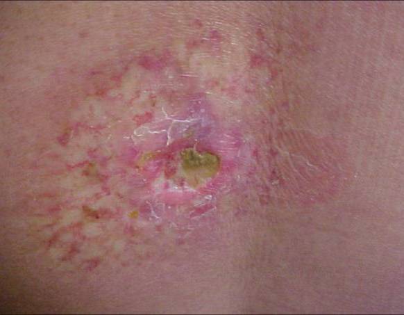
CLINICAL HISTORY: Our patient was 63-year-old caucasian man who presented to us in June 2006 for evaluation of an eight-year-old asymptomatic lesion on the back. He denied a history of radiation exposure or any trauma. There was a questionable history of a spider bite. A prior biopsy taken by another dermatologist during 2005 left a crusted non-healing ulceration. No resolution or improvement with topical steroids. The patient was switched to tacrolimus ointment twice daily and NbUVb three times weekly without improvement. Due to the complexity of the lesion, the patient’s history was reviewed again.
Review of the Past Medical History revealed extensive coronary artery disease, AAA, Diabetes Mellitus Type II, GERD, HTN, and hypercholesterolemia. A review of the Past Surgical History revealed cardiac stents placed in 1997, 1998, 2000, 2002, 2004, 2005 and another planned during 2006. There was a history of complex cardiac catheterization in 1998 and that the procedure time over four hours to perform. The patient recalled that the lesion first appeared a few weeks after the aforementioned difficult catheterization.
PHYSICAL EXAM:
85×55 mm well-demarcated, partially crusted, indurated, hypopigmented area with epidermal atrophy and telangiectasias.
LABORATORY TESTS: N/A
DERMATOHISTOPATHOLOGY:
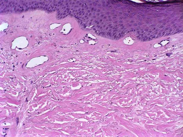
Initial report revealed an irregular epidermis, homogenization of the papillary dermis, and Sclerosis throughout the dermis with thickening of the dermis and compression of appendages with a few perivascular leukocytes consistent with morphea.
After reviewing the patient’s cardiac history, the case was discussed again with the dermatopathologist. In addition to dermal sclerosis, telangiectasias and atypical fibroblasts were seen.

DIFFERENTIAL DIAGNOSIS:
1. Morphea
2. Radiation Dermatitis
3. Necrobiosis Lipoidica


