CORRECT DIAGNOSIS:
Porphyria Cutaneous Tarda
DISCUSSION:
Porphyria Cutanea Tarda (PCT) is the most common type of porphyria [2, 3]. It is due to a deficiency in the enzyme uroporphyrinogen decarboxylase, leading to a buildup of porphyrins in the liver [4]. Most patients have an acquired form of the disease, but there are patients who possess an autosomal dominant deficiency of the enzyme UPD and a third group of patients with an associated exposure to hepatotoxins.
PCT is considered to be one of the hepatic porphyrias. When the disease becomes active, large amounts of porphyrins build up in the liver. When these increased porphyrins are activated by UV light in the Soret band (400-410 nm) they become unstable and create reactive oxygen species. These free radicals lead to damage that is clinically seen [5]. PCT can become active when environmental factors such as alcoholism, hepatitis C virus, estrogen use, HIV, and iron overload combine to cause the deficiency of uroporphyrinogen decarboxylase in the liver. Additionally, 20% of PCT patients are homozygous for the C282Y mutation that causes Hemochromatosis [5].
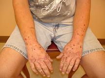
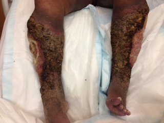
Skin lesions of PCT are characterized by bullae formation found on sun-exposed skin. The lesions tend to rupture easily, leaving behind erosions and shallow ulcers on otherwise non-erythematous skin. Lesions tend to heal with scarring, dyspigmentation, and milia formation. Patients tend to notice skin fragility with easy bruising and tearing, and hypertrichosis can often be found overlying the cheeks and temples. The diagnosis is often strongly suggested due to clinical findings alone. A useful office procedure that can be an easy confirmational test is the use of a Wood’s light exam of a random urine specimen, revealing a coral-red fluorescence. Further confirmatory studies would be increased urinary and fecal uroporphyrins, and slightly elevated fecal coproporphyrin and protoporphyrin levels. A skin biopsy would show subepidermal bullae with dermal papillae arising irregularly from the base of the split in the classic “festooning” pattern.
TREATMENT:
Treatment of acute porphyria should begin first with the removal of all potential precipitating agents, such as alcohol, medications and iron ingestions, and the treatment of underlying HCV infection if it exists. Sun protection in the form of barrier sunscreens and protective clothing is a must. Hydroxychloroquine in a low dose (200mg twice weekly) has also been recommended. Phlebotomy, however, is the treatment of choice for most patients, which alone or in combination with hydroxychloroquine may provide clinical remission.5 Uroporphyrinogen decarboxylase is inhibited by iron, so the removal of iron stores through phlebotomy may allow recovery of the diminished enzyme activity. For the rare patient who does not respond to the above treatments, desferrioxamine and erythropoietin treatment may offer an alternative treatment.
Our patient was treated with Plaquenil 200mg biweekly and phlebotomy weekly. His symptoms improved significantly after two months of therapy. His hepatitis C was managed with Ribavirin and Pegusus. He was encouraged to get immunization for Hep A & B series due to his Hep C infection. He was counseled to quit drinking alcohol and smoking and to use sunscreens regularly with SPF >30.
REFERENCES:
Bhawan, J., Sau, P., & Byers, H. R. (2004). Dermatology interactive atlas (ver. 5).
Elder, G. H. (1998). Porphyria cutanea tarda. Seminars in Liver Disease, 18, 67–75.
Bonkovsky, H. L., & Barnard, G. F. (2000). The porphyrias. Current Treatment Options in Gastroenterology, 3, 487–500.
Bonkovsky, L., Poh-Fitzpatrick, M., Pimstone, N., Obando, J., Di Bisceglie, A., Tattrie, C., et al. (1998). Porphyria cutanea tarda, hepatitis C, and HFE gene mutations in North America. Hepatology, 27, 1661–1669.
Bolognia, J. L., Jorizzo, J. L., Rapini, R. P., et al. (2008). Bolognia – Dermatology (2nd ed.). Spain: Elsevier.
Mascaro, J. M. (2003). Phlebotomy in sporadic porphyria cutanea tarda. Archives of Dermatology, 139, 343–345.


