Presenter: Reagan Anderson, DO
Dermatology Program: Oakwood Southshore Medical Center
Program Director: Steven Grekin, DO
Submitted on: June 20, 2008
CHIEF COMPLAINT: Masses on left jaw, right hand, and left ankle which have been progressively and symmetrically enlarging x 4 years
CLINICAL HISTORY: 6-year-old white female presents to the clinic with masses on left jaw, right hand, and left ankle which have been progressively and symmetrically enlarging for the last 4 years. She is asymptomatic and lesions do not interfere with daily life except for having to buy different sized shoes. So far, cheek and tongue lesions do not interfere with eating or swallowing and do not increase in size when illnesses are present. The patient was initially seen by multiple providers for “excess skin” on her right hand and left foot. Consultation at 3 years of age to Genetic and Metabolic Disorders at the Detroit Medical Center by Orthopedics was not conclusive but a diagnosis of neurofibromatosis (NF) type 1 was entertained. MRI of the left foot was performed at 3 years of age which was read as a likely venous or lymphatic structure. Follow-up with ultrasound was recommended by radiology but not performed. The patient was sent to Ophthalmology and had a normal examination.
Other information: The patient was borne full-term to a then 33-year-old G2P1 who reports a normal pregnancy with no complications and no known exposures to teratogens, illegal drugs, or radiation. The patient has attained all developmental milestones and is completely age-appropriate. Parents report no behavioral problems. The patient is otherwise in good health with no known illnesses or conditions other than the soft tissue masses. Family history is non-contributory except for a maternal first cousin once removed from the patient who has been diagnosed with NF type 1.
PHYSICAL EXAM:
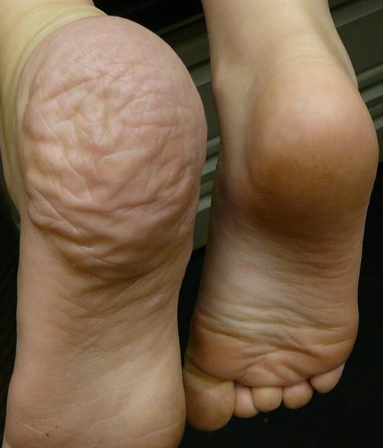
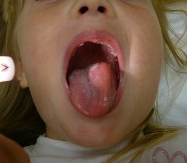
The left face has a prominent tumorous mass which extends into the buccal mucosal. Noted asymmetry of the left side of her tongue due to a soft-tissue mass is present. The right hypothenar eminence has a soft-tissue mass which does not seem adherent to underlying tendinous structures. The left ankle has a similar soft-tissue mass which encompasses most of the heel. No bruits or hypertrichosis are associated with the lesions. Additionally, the patient has multiple café au lait spots on her legs, back, and arm. Some are larger than 5mm. No other abnormalities are noted on physical examination.

LABORATORY TESTS:
Ophthalmology and skeletal x-rays (at 3 years of age). Spinal X-ray at 3 years of age showed levoconvex curvature which was likely positional in nature.
Left foot MRI at 3 years of age revealed a likely venous or lymphatic structure.
MRI of hand at 6 years of age revealed a likely venous or lymphatic structure.
Ultrasound performed at 6 years of age: left submandibular region and right hand are likely lymphatic malformations. Embolization was suggested as a possible treatment. The left foot has a more “diffuse-type of lymphatic malformation.”
DERMATOHISTOPATHOLOGY:
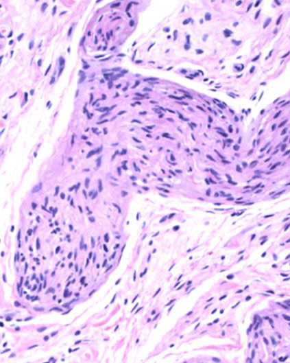
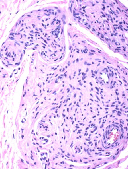
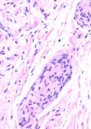
Two elliptical biopsies were submitted, one from the mass on the right hand and one from the left ankle. These showed “skin with hyperkeratosis with a diffuse proliferation of elongated cells in a myxoid background separated by fibrous septae. The plexiform arrangement is noted in the deep portion. No atypical changes are seen. Both lesions reach the margins of excision. No lymphatic malformation is identified.”
DIFFERENTIAL DIAGNOSIS:
1. Proteus Syndrome
2. Blue Rubber Bleb Nevus Syndrome
3. Maffucci’s Syndrome
4. Isolated Venous Malformation
5. Neurofibromatosis Type 1


