Presenter: Aaron Bruce, D.O., Roger Sica, D.O., Lyubov Avshalumova, D.O., Johnny Gurgen, D.O., Risa Ross, D.O., Rachel Epstein, D.O., Jessica Flowers, D.O., David Judy, D.O.
Dermatology Program: Nova Southeastern, Largo Medical Center, Sun Coast Hospital
Program Director: Richard Miller DO, FAOCD
Submitted on: August 27, 2008
CHIEF COMPLAINT: “Blisters on arms and legs”
CLINICAL HISTORY: We present a 50 y/o Caucasian female with a new onset of blisters on her thighs, arms, and axilla. Pt has a known history of Churg-Strauss Syndrome and states that she developed these blisters while on a prednisone taper. Pt denies any previous history of skin disease. She does state that these blisters become very irritated and painful at times. Pt denies oral lesions and constitutional symptoms. She denies starting, changing dosages, and frequency of any medications.
Pt has a known history of Churg-Strauss which is currently being managed with prednisone 2.5 mg and 5 mg alternating daily. Other past medical histories are significant for MVP and HTN. Pt denies tobacco, EtOH, and recreational drug use. NKDA. Medications include Fluticasone/Salmeterol inhaled, Triamcinolone nasal spray, Ramipril, ASA, Carvedilol, Digoxin, Spironolactone, Alprazolam, and Prednisone. Unremarkable family history.
PHYSICAL EXAM:
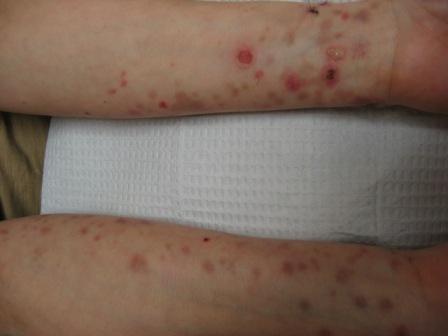
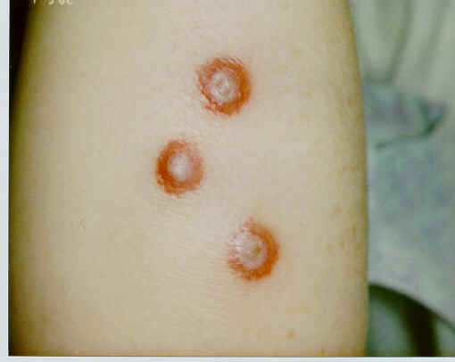
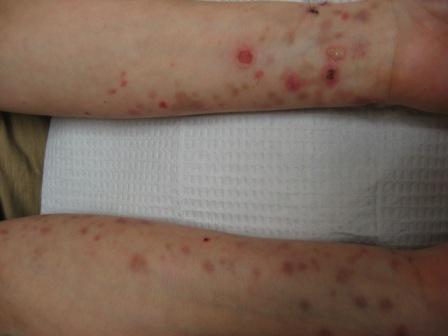
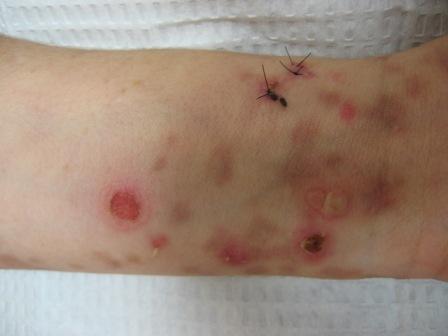
Examination of the skin reveals multiple tense bullae with surrounding erythema on the volar wrists bilaterally and extensor forearms. There are scattered hyperpigmented patches noted on bilateral arms and thighs. There are no oral lesions present.
LABORATORY TESTS:
CBC- WNL
CMP- WNL except slightly elevated potassium (5.5 mmol/L )
G6PD- WNL
TPMT- 14.6 U/mL RBC (range: 15.1-26.4 U/mL RBC)
IgA and IgG- WNL
ANA- negative
DERMATOHISTOPATHOLOGY:
Two 4 mm punch biopsies were performed. Both from the volar wrists.
H&E- subepidermal blister with increased polymorphonuclear leukocytes and eosinophils
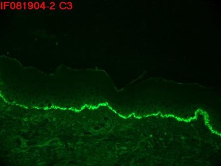
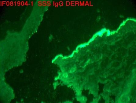
Direct IF- thick meshy linear IgG and much weaker C3 deposition along the epidermal basement membrane zone. After salt-splitting this specimen the thick linear IgG and Ce deposition was located only at the dermal side of the artificially induced blister. There is no IgA, IgM, C5b-9, or fibrinogen deposition observed in this specimen.
DIFFERENTIAL DIAGNOSIS:
1. Epidermolysis Bullosa Acquisita
2. Bullous Pemphigoid
3. Bullous Drug Eruption
4. Linear IgA Disease
5. Dermatitis Herpetiformis

