CORRECT DIAGNOSIS:
Epidermal Nevus Syndrome
DISCUSSION:
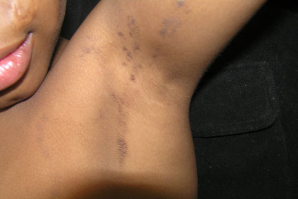
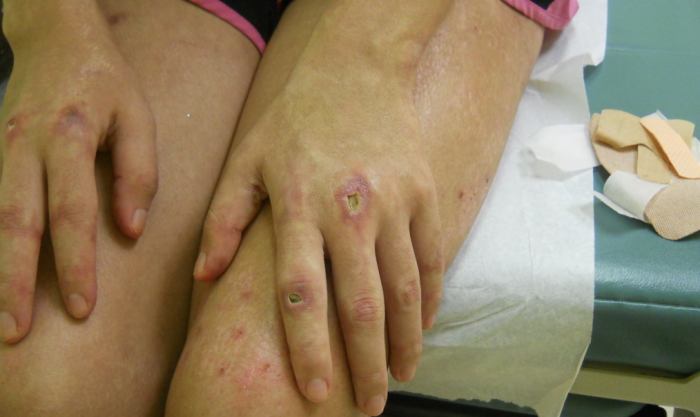
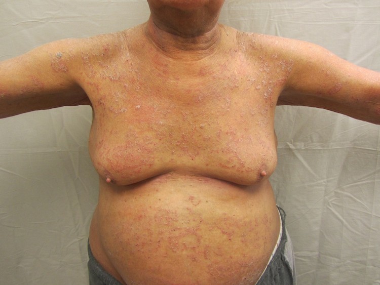
Epidermal Nevus Syndrome also is known as inflammatory linear verrucous epidermal nevus (ILVEN) syndrome is a rare neurocutaneous disorder characterized by specific skin lesions with significant involvement of the nervous, ophthalmologic, and skeletal systems. It represents a dysembryogenesis with both ectodermal and mesodermal malformations occurring in a regional pattern.
There are four clinical variants of this syndrome
-Linear Sebaceous Nevus(LSN)
-Linear nevus comedonicus (NC)
-Linear epidermal nevus (LEN)
-Inflammatory linear verrucous epidermal nevus (ILVEN).
ILVEN is a linear, persistent, pruritic plaque, usually first noted on a limb in early childhood. ILVEN is characterized by tiny, discrete, erythematous, slightly warty papules, which tend to coalesce in a linear formation.
Other key features of this syndrome are involvement of central nervous system characterized by mental retardation, Seizures, hemiparesis, paraparesis, sensorineural deafness, cerebral hemangiomas. Seizures are reported in 75% of patients and the morphology of the seizures varies from infantile spasms or focal motor seizures to the generalized tonic or tonic-clonic seizures. In some children, seizures are drug-resistant and may result in progressive mental retardation.
Skeletal involvement is associated with the location of nevi and may include hemihypertrohpy, kyphoscoliosis, Vitamin D-resistant rickets, and foot/ankle deformities.
Eye involvement is due to extension of nevus to the lid and bulbar conjunctiva which may lead to corneal opacity, blindness, or nystagmus.
In rare cases, variety of neoplasms are associated with this syndrome which includes Wilm’s tumor, astrocytoma, rhabdomyosarcoma, syringocystadenoma papilliferum, and salivary gland adenocarcinoma.
Laboratory data:
-Skin biopsy: Reveal’s hyperkeratosis, papillomatosis, and acanthosis with rete ridge elongation in a psoriasiform pattern.
-EEG findings are abnormal in approximately 90% of patients. In almost all patients who had focal paroxysmal electroencephalographic abnormalities, the epileptiform focus was ipsilateral to the major skin lesions.
-MRIs can be used to evaluate intracranial involvement. MRIs may show cerebral atrophy, dilated ventricles, hemimegalencephaly, pachygyria, or enlarged white matter.
-Serum calcium/phosphorus. Patients with epidermal nevus syndrome have hypophosphatamia and excision of the nevus or supplemental phosphate with vitamin D may help halt the progression of bone and CNS changes.
TREATMENT:
Topical steroids, topical lactic acid in propylene glycol, surgical removal, dermabrasion, Co2 laser. Also referral to neurologist, ophthalmologist, and orthopedics to manage other associated problems.
REFERENCES:
Rogers, M., McCrossin, I., & Commens, C. (1989). Epidermal nevi and the epidermal nevus syndrome: A review of 131 cases. Journal of the American Academy of Dermatology, 20(3), 476-488. https://doi.org/10.1016/S0190-9622(89)70084-6
Altman, J., & Mehregan, A. H. (1971). Inflammatory linear verrucose epidermal nevus. Archives of Dermatology, 104(4), 385-389. https://doi.org/10.1001/archderm.1971.04010040075005
Fink-Puches, R., Soyer, H. P., Pierer, G., Kerl, H., & Happle, R. (1997). Systematized inflammatory epidermal nevus with symmetrical involvement: An unusual case of CHILD syndrome? Journal of the American Academy of Dermatology, 36(5 Pt 2), 823-826. https://doi.org/10.1016/S0190-9622(97)80462-2
Golitz, L. E., & Weston, W. L. (1979). Inflammatory linear verrucous epidermal nevus: Association with epidermal nevus syndrome. Archives of Dermatology, 115(10), 1208-1209. https://doi.org/10.1001/archderm.1979.01630060060009


