Presenter: Jonathan Cleaver D.O., Peter Knabel D.O
Dermatology Program: Northeast Regional Medical Center
Program Director: Lloyd Cleaver D.O
Submitted on: June 5, 2011
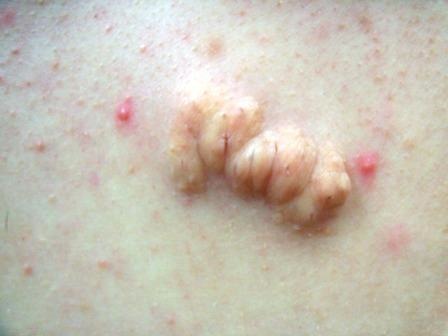
CHIEF COMPLAINT: Soft asymptomatic lesion on the upper mid-back
CLINICAL HISTORY: A 16-year-old well developed female presents to the clinic for evaluation of a lesion on her back that has been present since she was an infant. The lesion has continued to increase in size as she has developed. The lesion is asymptomatic and the patient denies tenderness, drainage, bleeding, or color change. No one else in the family has a similar lesion. She has been healthy since birth and there is no significant family medical history reported. The patient is on no medication. The lesion has been manipulated by the mother multiple times trying to express material from the lesion. She has been unsuccessful at these attempts.
PHYSICAL EXAM:
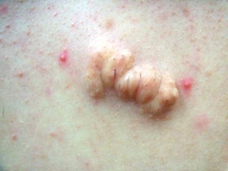
A well-defined 3cm x 1.5cm flesh-colored papillomatous plaque on the upper mid-back. There were few surface comedones scattered throughout the lesion. The lesion had a soft rubbery consistency and was not fixed to deeper structures.

LABORATORY TESTS: N/A
DERMATOHISTOPATHOLOGY:
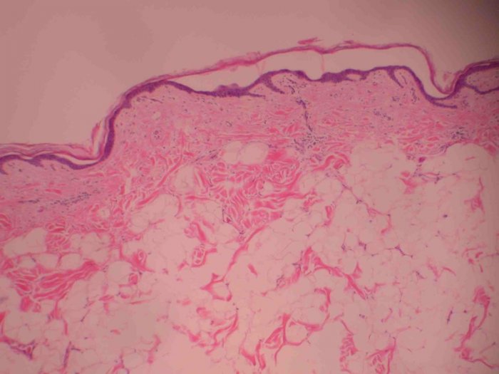
Histological examination demonstrated slight papillomatosis as well as borderline basal layer pigmentation. The underlying dermis shows the usual fibrocollagenous tissue. Mature adipose tissue is present within the dermis between collagen bundles. No spindle cell lesion or prominent inflammation is identified.
DIFFERENTIAL DIAGNOSIS:
1. Plexiform Neurofibroma
2. Connective Tissue Nevus
3. Penduculated lipofibromas
4. Nevus lipomatosus superficialis
5. Focal dermal hypoplasia

