CORRECT DIAGNOSIS:
Autoimmune Progesterone Dermatitis
DISCUSSION:
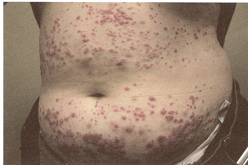
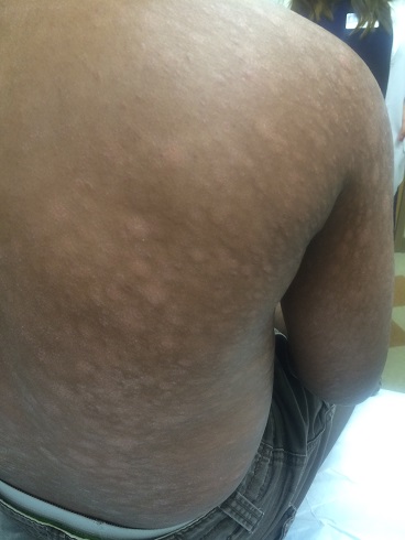
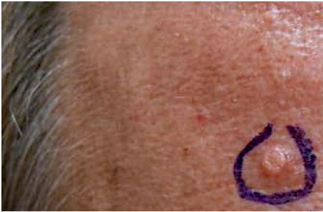
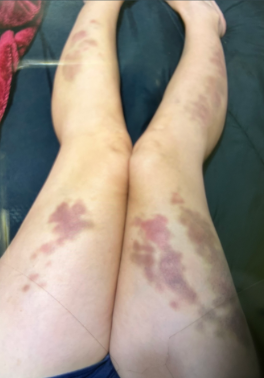
Autoimmune progesterone dermatitis (APD) is classically described as a cyclical cutaneous eruption, occurring or worsening during the luteal phase of the menstrual cycle when progesterone levels are elevated. APD typically occurs in women between the ages of 12-44. Patients with APD often present with non-specific symptoms for 5-6 years before diagnosis. The cutaneous eruption can vary in morphology. The lesions may be urticarial, papular, papulovesicular, eczematous, or erythema-multiforme like. Mucosal surfaces may also be involved. The eruption has no predilection for any specific body site. Symptoms generally appear or worsen within 10 days prior to menses, and resolve or begin to improve with the onset of menses. Pruritus is typically reported, but the eruption may also be asymptomatic.
The diagnosis of APD is typically made on the clinical presentation and history of a cyclical eruption around menses. Improvement after ovulation suppression therapy or worsening after removal of suppression supports the diagnosis. A positive intra-dermal progesterone test performed with a small amount of progesterone suspension 50mg/ml can also support the diagnosis. The reaction can take anywhere between 30 minutes to 96 hours to become positive. Histological features of suspected APD vary widely and there are no clear-cut unifying features for the APD cases reviewed. A biopsy may be needed to distinguish APD from other dermatoses.
The exact pathogenesis of APD is poorly understood. It is generally agreed to represent an autoimmune condition in which the body forms a response to endogenous progesterone, manifesting itself clinically when progesterone levels are elevated. The highest levels of progesterone are typical during the luteal, or secretory, phase of the menstrual cycle.
TREATMENT:
The treatment for APD is primarily suppression of ovulation through oral conjugated estrogen therapy (oral contraceptives). Antihistamines, systemic steroids, and topical steroids are options in patients unable to take oral conjugated estrogen therapy or as an adjuvant therapy along with the conjugated estrogens. The prognosis is generally favorable as the treatment is typically successful.
The patient is controlled on a Medrol dose pack. She takes 1-2 tablets as needed during the luteal phase of her menstrual cycle. She has irregular cycles and so the cutaneous eruption is not consistent. She is not a candidate for oral hormone replacement therapy due to her age and the use of cigarettes. She also applies topical mometasone cream 0.1% as needed.
REFERENCES:
Chawla, S. V., Quirk, C., Sondheimer, S. J., & James, W. D. (2009). Autoimmune progesterone dermatitis. Arch Dermatol, 145(3), 341-342. PMID: 19255317
Herzberg, A. J., Strohmeyer, C. R., & Cirillo-Hyland, V. A. (1995). Autoimmune progesterone dermatitis. J Am Acad Dermatol, 32(2), 333-338. PMID: 7865603
Stranahan, D., Rausch, D., Deng, A., & Gaspari, A. (2006). The role of intradermal skin testing and patch testing in the diagnosis of autoimmune progesterone dermatitis. Dermatitis, 17(1), 39-42. PMID: 16510701


